Following a medical imaging test (ultrasound, MRI, PET scan), did the doctor tell you about a “spot”?
In the medical field, and more particularly in oncologythe presence of a stain during an imaging test – such as x-rays, mammograms, ultrasounds, CT scans or MRIs – may be anxiety inducing for the patient. “These anomalies may be discovered during a screening examination, particularly breast cancer screening, as part of an oncological care follow-up course, or incidentally when carrying out examinations as part of a other illness” specifies Dr Guillaume Le Bihan, oncologist-radiotherapist. This is what it could mean.
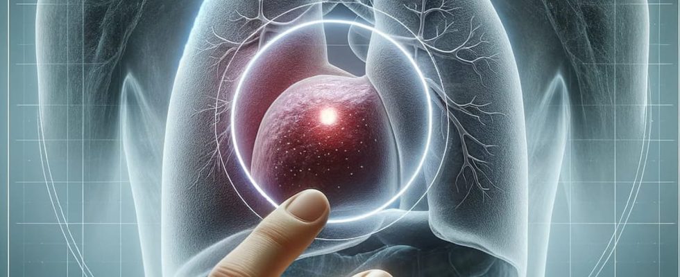
A spot in the breast, lung, liver or brain?
A “stain” can be detected at:
- Of the titssometimes indicating nodules, cysts, tumors, whether benign (non-cancerous) or malignant (cancerous)…
- Of the lungs, taking the form of pulmonary nodules. “They can be benign or malignant. comments Dr Guillaume Le Bihan, oncologist-radiotherapist
- Of the bone : Bone spots may indicate abnormalities such as injuries, fractures, bone infections (osteomyelitis), or bone metastases from a primary cancer that initially appeared elsewhere in the body.
- Of liver. “They can then be associated with liver masses, such as cysts, hepatomas (liver tumors) or metastases of cancers from other organs.”
- Of brain : “Brain spots detected using MRI or brain CT may be linked to primary brain tumors, metastases, vascular lesions, or other abnormalities.”
The spots can also appear on other organs such as the kidneysabdominal organs, gastrointestinal tract…
What is a stain? Cancer ?
“It is a generic term which does not systematically mean cancer“ reassures Dr Guillaume Le Bihan. There may be other anomalies such as:
► Benign lesions: certain spots can be cysts, nodules or other formations which are not cancerous. “For example, a simple fluid cyst in the liver or a benign nodule in the breast may appear as a blemish on a medical image.”
► Fibroids (also called leiomyoma or fibromyoma): these are non-cancerous tumors which develop, for example, in the muscular tissue of the uterus. “These tumors are composed of smooth muscle cells and connective tissue.”
► Inflammatory lesions such as granulomas. “They can take the form of a mass on images, particularly in the lungs.”
The detection of a stain on a medical image can also be interpreted as a recurrence of cancer without necessarily being a metastasis.
The term “metastasis” refers to the spread of cancer from its original site (the primary site) to other parts of the body. “A new mass, if malignant, can nevertheless be interpreted as a new cancer pathology and not only as a metastatic evolution of a primary cancer, specifies Dr Le Bihan. A pathological analysis by biopsy is sometimes necessary to distinguish between these two distinct situations.”
What to do ?
“Depending on the size, radiological appearance of the mass and the patient’s clinical assessment, additional investigations, such as biopsies, will be proposed. This will help determine whether the abnormalities are benign (non-cancerous) or malignant (cancerous).” A stain may also require simple second check radiological a few months later if the first x-ray is not worrying. He is crucial that any concerns regarding a spot detected on a medical image be discussed with a healthcare professionalpreferably an oncologist, radiologist or organ specialist. “Only a physician can correctly interpret imaging test results and recommend next steps based on the clinical situation.”
Thanks to Dr Guillaume Le Bihan, Oncologist-radiotherapist at the Onco-Radiotherapy center of the Polyclinique Bordeaux Nord Aquitaine (33) and Polyclinique Bordeaux Rive Droite (33)