Published on
Updated
Reading 2 min.
Marie Lanen
Head of parenting section (baby, pregnancy, family)

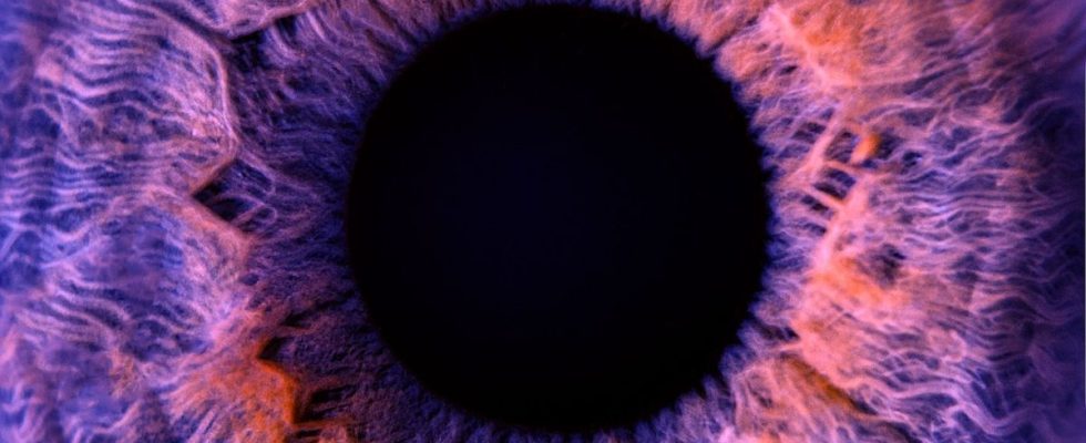
A Franco-Swiss research team has succeeded in making an entire human eye completely transparent. They managed to remove the opacity of the latter to study it in depth under the microscope. A technical feat that will make it possible to study eye diseases and their mechanisms.
The work of the Franco-Swiss research team was published on October 10 in the journal Communications Biology. It took 7 years of work for Marie Darche (research engineer and member of Professor Michel Paques’ team at the Quinze-Vingts hospital and the Paris Vision Institute), and the institute’s researchers. Wyss in Geneva to achieve this world first: the transparization (making transparent) of an entire human eye.
How to make a human eye transparent?
In an article published on their site, the Quinze-Vingts hospital mentions that “until now, the human eye was considered the organ most resistant to the transparency technique due to its complexity, its pigmentation and the fragility of its retina. This technique transforms an initially opaque biological sample into a transparent sample. It is then possible to see through it and, thanks to fluorescent antibody markings and microscopes specialized in large samples (light sheet microscopes), to visualize in 3D the organization of the cells and structures of the organ.“. The two research teams shared the work: Marie Darche took care of the “clearing” stages, through different manipulations (a succession of baths in organic solvents associated with marking with fluorescent antibodies). The Swiss team used a light sheet microscope (MesoSPIM) to obtain previously unseen images on the human eye.
Please note: the eyes come from deceased American donors. In fact, French legislation does not allow this specific type of donation (the French eye bank only receives corneas).
Better understand the evolution of eye diseases
The main objective of this major advance is to understand the evolution of eye diseases using high-resolution imaging. Thus, the research team has already planned to receive other eyeballs, this time pathological: suffering from AMD (age-related macular degeneration), glaucoma and high myopia in order to better understand the mechanisms of these diseases. . “A bank of healthy and pathological eyes imaged as the work progresses will be shared on request with international collaborators. A single sample can thus help answer multiple scientific questions and enrich various research projects. This association of medical, interdisciplinary and international experiences and knowledge centered around the same image bank will help us in the future to better understand the evolution of ocular diseases, to identify potential therapeutic targets or to evaluate the effectiveness innovative treatments” can we read on the website of the Quinze-Vingts hospital.
To go further, the National Hospital of 15-20 aims to develop the first French light sheet microscope entirely dedicated to the imaging of human samples in order to push the limits of scientific knowledge for the benefit of patients around the world. entire.
