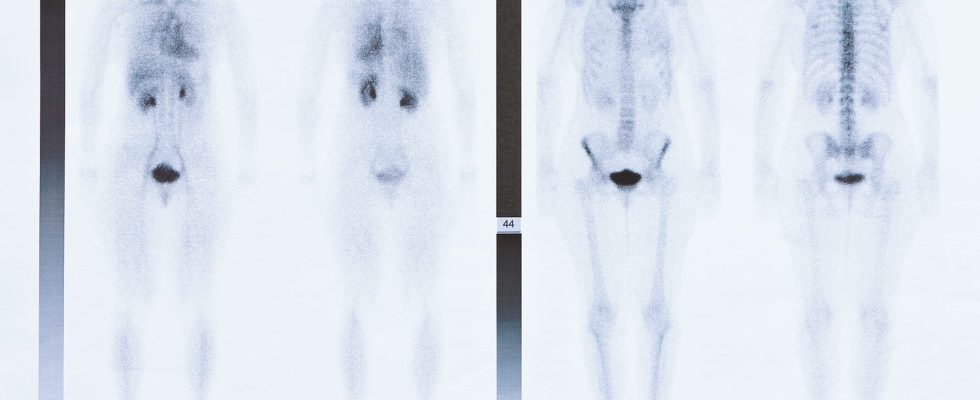
Scintigraphy is a medical imaging technique that uses radioactive substances to produce images. We can explore the lungs, the heart, the bones, the kidneys, the liver, the thyroid…
Definition: what is a scintigraphy?
scintigraphy East a medical imaging technique performed only in nuclear medicine departments. It consists injecting the patient with a radioactive substance into an organ or tissue. The radiation emitted by the substance will settle on certain areas, and draw a visual map of the area to be explored. There are many types of scintigraphy : bone, pulmonary, cardiac scintigraphy, etc.
What is a scintigraphy used for?
This reviewsafe due to very low dose of radiation, can highlight many conditions. It is used in particular:
- to look for bone metastases in the cancer spread assessment,
- to look for a pulmonary embolism,
- diagnose or follow the evolution of a bone pathology : Paget’s disease, myeloma, stress fracture, trauma, bone tumor
- perform a bone assessment in case of hypercalcemia in case of bone and joint infection
- detect heart, lung, kidney or thyroid dysfunction
What is a bone scan used for?
Bone scintigraphy is a medical imaging examination that involves injecting a substance (marker) into the body and observing the reaction produced. It is particularly indicated to identify metastases in cancer patients. Once injected, the marker binds to abnormally active cells such as cancer cells. Thus, it is possible to locate a metastasis from the reconstructed image. The scintigraphy is practiced less and less since the appearance of MRI.
What is a brain scan used for?
Brain scintigraphy is an examination carried out using a radioactive product. It makes it possible to visualize cerebral activity by highlighting the areas of the brain which are more irrigated when they are solicited. This examination is of interest in precisely locating areas of hyperactivity in case of epilepsy notably. The examination lasts approximately one hour and only requires an injection or an infusion.
What is a myocardial scintigraphy used for?
Myocardial scintigraphy is an examination indicated in cardiology in case of suspected coronary disease. It consists in studying the functioning of the heart, in particular its vascularization. This examination is carried out in addition to a stress test carried out on a bicycle or a treadmill. Myocardial scintigraphy is especially useful in patients with risk factors for coronary artery disease (diabetes, smoking, high blood pressure, high cholesterol).
What is a lung scan used for?
A lung scintigraphy is a medical imaging examination to observe the return of venous blood and/or ventilation. To assess venous circulation, radioactive particles are injected into a vein and these are observed using a camera capable of detecting them. Normally, particle-laden blood should go to both lungs. Ventilation lung scintigraphy, on the other hand, makes it possible to ensure lungs are adequately ventilated. In this case, it is a radioactive gas which is inhaled and analyzed with the gamma camera.
What is a kidney scan used for?
Kidney scintigraphy is a test to visualize kidney activity, detect possible malformations of the kidney or elimination pathways (urethra, bladder). It is performed by injecting a radioactive product into the venous network. The examination will make it possible to see this product being collected at the level of the kidney then being eliminated by the natural ways. It thus highlights obstacles to the elimination, reflux, malformations or scarring and assesses kidney function.
In case of late menstruation or doubt about a pregnancy, it is best not to practice it.
What is a thyroid scan used for?
The thyroid scintigraphy is a medical examination of the thyroid which allows a functional analysis of this gland. The scintigraphy consists of administer slightly modified iodine, which the thyroid will fix and the secretion of labeled iodine makes it possible to locate an overactive or underactive zone. If the substance is evenly distributed, it means that the thyroid is working properly. The scintigraphy of the thyroid is associated with biological examinations, in particular the dosage of TSH, and a thyroid ultrasound.
No preparation is required before a scan. Before the examination, the medical staff injects a product slightly radioactive in a vein in the arm or by gas inhalation in case of ventilation lung scintigraphy. Then the camera saves the photos. This examination is completely painless. Depending on the indication, the first images are taken immediately after the injection, and the following 2 to 4 hours later to give the product time to spread through the body. After the examination, it is advisable to drink plenty of water to accelerate the elimination of the product.
Is a scan dangerous?
Scintigraphy does not present any danger. There amount of radioactive product injected is small and does not expose the patient to health risks. Allergies exist but are exceptional. Driving a vehicle is possible after a scan. However, in case of late period or doubt about a pregnancy, it is best not to practice it. Scintigraphy is contraindicated in pregnant or breastfeeding women.
