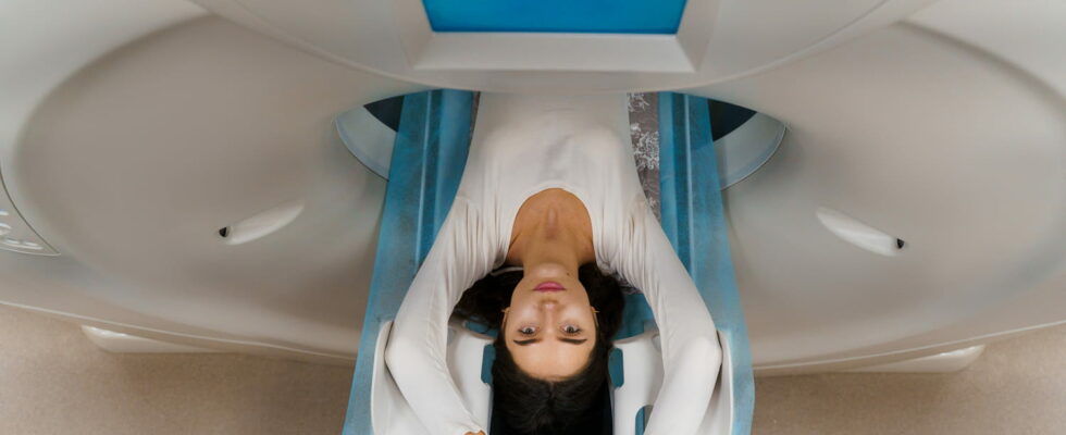
The scanner is a medical imaging examination, also called “computed tomography”, which has the advantage of analyzing the organs of the human body “in section” in a very precise way but also the tissues, bones, joints… Unrolled.
The scanner formerly called “computed tomography” is a medical imaging technique which allows toobserve and study the organs of the human body by visualizing them fine cuts, more precisely than other imaging techniques such as x-ray and of theultrasound.
What is a CT scan in medicine?
The scanner allows take precise pictures of the organs of the human body (brain, liver, lungs, pancreas, spinal cord, bones, joints, etc.). The technique used is close to radiography: the device emits X-rays which revolve around the patient and therefore make it possible to obtain the image not in 2D but in 3D volume of the entire inspected area. THE Scanner images are “cutaway”. THE images will be more or less contrasted depending on the tissues and organs crossed. The scanner can be performed on a specific part of the body, or on the whole body, in particular in the event of a search for metastases in the assessment of cancer extension, or of significant trauma.
How long does a scan take?
The exam itself lasts between 5 and 20 minutes with an average of 10 minutes. For the complete examination (reception, examination, return of results), it generally takes 1 hour. The delay is longer when the scanner requires the injection of a contrast product.
What is the scanner used for?
The exam gives very precise information on the organ(s) studied (size, shape, structure, presence of abnormal lesions, etc.). It thus allows a very precise vision of all the organs. But also of highlight internal lesions within certain organs, including the density will make it possible to characterize them and deduce their nature (especially in the diagnosis of cancer). The device defines the volume, the consistency, their extension to the nearby organs as well as their contours. The images can be multiplied according to different planes to obtain a virtual sectional view and a reconstruction of extremely detailed images. Can be visualized in this way: the thorax, the brain, the abdomen, the bones…
Can the scanner detect cancer?
Yes, the scanner is an examination that can help in the diagnosis of cancer but also “for the evaluation of the effectiveness of a treatment or for the follow-up after the end of the treatments” explains The League Against Cancer.
Types of medical scanners
What are the CT indications?
The scanner allows diagnose certain pathologies, or of monitor the evolution of tumors during treatment to define their volume and extent (search for metastases). It can be done to monitor a possible relapse. It is recommended for looking for abnormalities that would not be visible on a standard X-ray, on an ultrasound or as an alternative to MRI. It allows further examination of the thorax (suspicion of infection, pulmonary embolism, etc.), abdomen, limbs (complex fractures, calcifications, etc.). Full body scan may be requested in accidents causing polytrauma. A brain scan may be requested in case of violent headaches, brutal or unusual. It can be used to find, for example, a malformation or an aneurysm.
Scanner with or without injection
The scan is performed with or without injection of one iodinated contrast agent. This product is necessary to observe the targeted organ more precisely and improve the quality of the images. It is given intravenously most often and can cause a feeling of warmth. It can leave a metallic taste in the mouth and sometimes nausea. When a contrast product has been injected, it is advisable to drink plenty after the examination, throughout the day (water, tea, coffee, juice, etc.) to eliminate it.
Any allergies to iodine must be reported
The patient is lying on an examination table and must remain still for the duration of the procedure (he is alone in the room). When the exam begins, the table advances in a tunnel-shaped machine that is open and shallow compared to the MRI where the tunnel can appear more impressive. The X-ray beam scans the affected area. A radiographer remains attentive during the examination to check that it is going well and the patient’s reactions.
Preparation before a scan
He is recommended to be on an empty stomach (or to take a light meal to avoid nausea or even vomiting afterwards) whether contrast solution is administered orally or rectally “but in general, you should not interrupt your current treatment and take your usual tablets the morning of the exam“notes Professor Vincent Vidal, specialist in interventional radiology at the Timone hospital in Marseille – AP-HM. Any allergies to iodine must be reported to health professionals. Taking an anti-allergy medication may be considered. For a scanner:
- no clothes with metal snaps or buttons
- no jewelry
- no piercings
- no metal bars or clips
Is a CT scan painful?
A CT scan is not painful. The injection of the contrast solution can cause a sensation of heat in the body.
How much does a medical scanner cost?
The cost of a CT scan varies from 30 euros to less than 100 euros, depending on the area inspected and whether or not the examination requires an injection of contrast solution. THE examination prices are set by the Health Insurance for professionals working in sector 1. They are reimbursed on the basis of 70% of the convention rate as part of the coordinated care pathway. When they exercise in sector 2, healthcare professionals can charge for the examination with excess fees.
Can you have a CT scan while pregnant?
The scanner is not not indicated for pregnant women : X-rays can be dangerous for the foetus. It is also necessary check the absence of renal insufficiency as well as the drugs taken by the patient, in particular certain oral antidiabetics which will need to be stopped for a short time after the scan.
Thanks to Pr Vincent Vidal, specialist in interventional radiology at the Timone hospital in Marseille – AP-HM
Source: Scanner or computed tomography, patient sheet, National Cancer Institute, September 2020.
