The uterus is the organ of gestation which belongs to the female reproductive system. What are the shapes of the uterus? What are the abnormalities and diseases of the uterus?
The uterus is a female reproductive system organ. It is the organ in which the embryo develops during a pregnancy. There are several forms of uterus (normal, anteverted, retroverted etc). There are malformations of the uterus (cloistered, contractile) which cause complications in women. What are the examinations of the uterus? What are the diseases of the uterus?
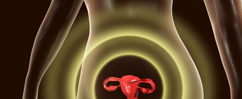
Definition: what is a uterus?
the uterus is a pear-shaped organ that is part of the female reproductive system. With each menstrual cycle, under the effect of female hormones, its mucous membrane (or endometrium) thickens to promote the eventual implantation of an embryo. When there is fertilization, the embryo lodges there (it’s here implantation) and grows there. The size of the uterus increases as the pregnancy progresses up to about 35 cm in height. If there is no fertilization, the mucous membrane is destroyed: these are the rules.
What are the different shapes of the uterus?
► The anteverted uterus. The uterus is normally anteverted, i.e. tilted forward. Its version angle is located in front of the axis formed between the navel and the coccyx. It then rests on the bladder.
► The retroverted uterus. “It’s about a anatomical variant generally harmlessexplains Dr. Christelle Besnard-Charvet, gynecologist. Instead of normally leaning forward, between the bladder and the rectum, it is positioned backwards, in line with the vagina and can press on the rectum. This positioning is usually asymptomatic, but can cause period pain felt in the back“. It does not prevent you from getting pregnant or carrying a pregnancy to term without risk.
► The artificial uterus corresponds to a hypothetical substitute organ which would allow an embryo to develop outside the woman’s body until it fully matures. This device ofectogenesis is often referred to as an extracorporeal pregnancy. Thus, the artificial uterus would have the function of allowing women with a damaged uterus to carry a pregnancy to term, outside their body. It could also increase the viability threshold of premature babies.
What are uterine malformations?
Certain congenital malformations can mark this organ. “Among them, the bicornuate and septate uterus, the absence of a uterus in some women or even too small uteri when the patient’s mother took distilbene“, says the gynecologist.
► The bicornuate uterusalso called didelphe uterus, is a congenital malformation: instead of being pear-shaped, it is heart-shaped, and smaller than normal in size. Although not serious and usually asymptomatic, the bicornuate uterus can affect the smooth running of a pregnancy. More rarely, the bicornuate uterus can be a cause of infertility.
► The septate uterus is a congenital malformation. It is characterized by the abnormal presence of a internal dividing wall. Thus, the uterus is divided into two distinct cavities. This separation can be partial or extend to the cervix. Note that among congenital uterine malformations, the septate uterus is the most common. “It can cause the same complications as the bicornuate uterus, namely miscarriage, premature delivery, breech presentation, growth retardationbut it is more rare, it all depends on the size of the partitionsays Dr. Besnard-Charvet. Surgery is possible, it is recommended when there is a complication: it consists of resecting the septum by natural means under hysteroscopic control“.
► Contractile uterus refers to a uterus that contracts abnormally before the end of the 9 months of pregnancy. Contractions of the uterus may or may not be painful. If the pregnant woman feels more than 10 contractions per 24 hours, she should consult her doctor to ensure that they are not “effective”, i.e. causing an opening of the cervix and a risk of premature delivery. To limit contractions of the uterus, rest will be advised to the future mother.
What are the examinations of the uterus?
► The cervico-vaginal smear. Free for all women between the ages of 25 and 65 under the cervical cancer screening, the cervico-vaginal smear is a painless examination: carried out by your gynecologist, it consists of removing superficial cells by gently rubbing them with a spatula or a small brush. If the cells taken during the smear are suspicious, additional examinations are carried out. Among them, a colposcopy, an examination of the cervix carried out using a microscope in the gynecologist’s office: it makes it possible to observe suspicious areas precisely thanks to the application of colored reagents making it possible to highlight the injured areas and to perform a biopsy more easily.
► Hysteroscopy. This examination allowsstudy the inner wall of the uterus using an endoscope. This accessory consists of a flexible tube equipped with an optical fiber. This examination makes it possible to look for polyps, signs of inflammation or infection, a malformation and a tumour. “It is recommended in infertility check-ups, in case of repeated miscarriage and heavy or frequent pain or bleeding, especially after menopause“, adds Dr. Besnard-Charvet. It allows all at the same time to detect anomalies, to take samples and to carry out small surgical gestures such as the resection of polyps or fibroids.
What are the diseases of the uterus?
► Uterine fibroids are small benign tumors, non-cancerous, which develop in the uterine muscle. Very frequent, they concern 40% of women. They usually have a round shape. They can be inside the organ (intra-uterine fibroid), or outside (extra-uterine fibroid) by simply being attached by a foot (pedunculated fibroid). Because they are very often asymptomatic, they can go unnoticed for many years. It is during a medical examination or an ultrasound that they are discovered. But some can cause inconvenience such as heavy bleeding during and outside of menstruation, pain during intercourse, frequent urination, episodes of constipation and a feeling of discomfort in the lower abdomen. Depending on its size, the fibroid can also impact fertility by preventing implantation from taking place.
► Cervical cancer is the 12th leading cause of cancer and the 12th leading cause of cancer death in women. Each year, approximately 3,000 new cases are discovered. It is related to a virus, human papillomavirus (HPV). Contamination mainly takes place during unprotected sexual intercourse. Its detection is based on the regular practice of a smear which, by taking cells from the cervix, allows analysis under a microscope. Treatment then consists of surgery, radiotherapy or chemotherapy. Since 2007, there has been a cervical cancer vaccine (Gardasil), reimbursed at 65% by Health Insurance, for young girls under 14 years old.
► A polyp is a benign growth. It can be single or multiple. Generally asymptomatic, it can sometimes be responsible for bleeding. They are discovered fortuitously during a routine gynecological consultation or a pelvic ultrasound. They may be responsible for pain, bleeding during and after menstrual periods, infertility and profuse genital discharge. Drug treatment based on progesterone may be effective on small polyps. For others, surgical removal will be recommended.
► endometriosis is manifested by violent pelvic pain, mainly during menstruation, and can even lead to infertility. It is characterized by the presence of endometrium outside the uterus, which is lodged on the cervix, fallopian tubes, ligaments, ovaries, peritoneum, vagina, vulva… Sometimes, this mucous membrane is also deposited on non-genital organs: bladder, appendix, colon, sigmoid… These segments of uterine lining follow the hormonal rhythm and produce blood every 28 days. “The clinical signs make it possible to suspect the diagnosis, in particular period pain, but also pain during intercourse“, says our expert. Ultrasound and especially pelvic MRI can confirm the diagnosis. The treatment is then based on the prescription of analgesics and anti-inflammatories to fight against the pain and hormonal treatments (continuous contraceptive) to put an end to the rules, and thus the pain and bleeding. In case of failure, surgery will be considered: the aim is to remove endometriosis lesions, usually performed by laparoscopy, causing endometriosis pain. As a last resort, the total removal of the uterus will be recommended.
► Removal of the uterus also called “hysterectomy”, it is a total or partial removal (it is possible to keep the cervix) of the uterus. It can be performed using several techniques, under general anesthesia: vaginally or abdominally. It is practiced in some cases of cancers of the genital tract that can affect the cervix, endometrium, lining of the uterine wall, or ovary. “It is only indicated in the event of pathology of the uterus causing symptoms: heavy periods linked to fibroids or polyps, cancer of the uterus, descent of organsexplains the gynecologist. It can also be conservative, that is to say that the intervention preserves the ovaries: in this case, if the woman was not menopausal before the intervention, the only difference is the cessation of the rules. Finally, it can be associated with removal of the ovaries, for example in the event of ovarian cancer, which will create a surgical menopause.“.
► Conization involves removing only part of the cervix to eliminate lesions that could develop into cancer. Each year in France, 25,000 women are affected. It is generally proposed after the realization of a smear having revealed the presence of pathological cells. Performed in the hospital, on an outpatient basis, under loco-regional anesthesia, it consists of remove a small portion of the cervix with a scalpel or laser. “If the conization is minimal, it does not interfere with fertility, nor the evolution of pregnancy“, specifies the specialist.
Thanks to Dr Christelle Besnard-Charvet, gynecologist.