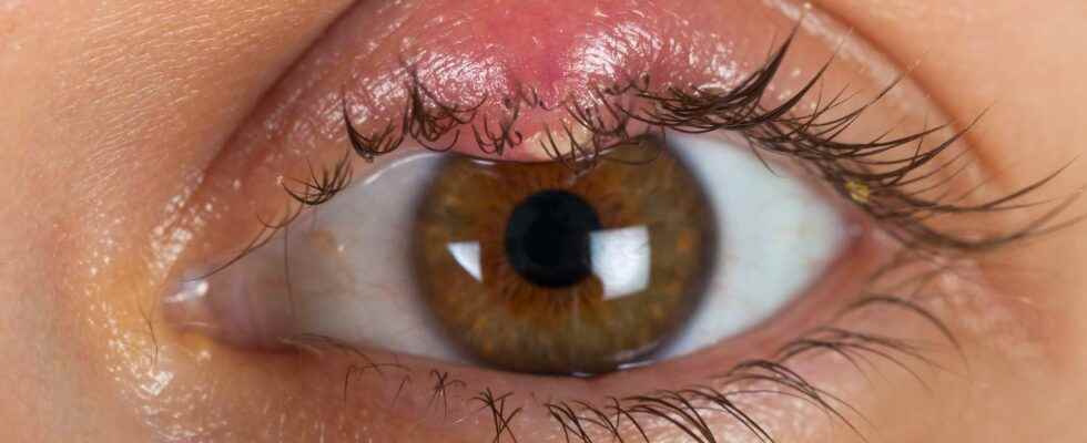
stye is a small infection that is located at the base of the eyelash of a eye. He is without gravity but can be painful and embarrassing. Usually it resolves after a few days on its own, but sometimes requires a visit to a doctor.
What is a stye?
The eyelashes, skin appendages located at the edge of the eyelids protect the eyes and are made up of a rod of keratinized cells implanted in the dermis inside a cavity: the follicle hairy. The latter contains nerve endings and accessory glands (sweat and sebaceous). The stye is the result of a bacterial infection, usually staphylococcus, which develops in this cavity. An abscess develops, with accumulation of pus and materials at the edge of the eyelid. The causes can be various: rubbing of the eyemakeup, wearing lentils and its occurrence is more common in people with diabetesof drought eyes, of dermatitis seborrheic or presenting with immune deficiency.
What are the symptoms of a stye and what treatment should be adopted?
A stye often begins with painful redness and swelling at the base of an eyelash that can lead to itching. The eye tears and becomes sensitive to lighta lump appears the size of a grain ofbarley, fills with pus and eventually pierces spontaneously after a few days. Benign but nonetheless embarrassing and painful, this condition recidivism frequently in the absence of antibiotic therapy. The treatment is based onapplication hot water compresses on the lesion.
The iris, the colored part of the eye The iris is the colored area of the eye, visible from the front. It is in fact the anterior part of the vascular tunic (choroid). The iris has an opening in its center: the pupil, round, which lets light through. The diameter of the pupil can vary, allowing more or less light to enter, depending on the ambient light. The iris contains a more or less abundant brown pigment. © Mattis2412, CC by-sa 3.0
The lateral rectus muscle rotates the eye The lateral rectus muscle of the eye rotates this organ around an axis, thus allowing the gaze to be directed outwards. © Patrick J. Lynch, CC by 2.5
Consultation with the ophthalmologist During an examination at the ophthalmologist, the professional uses different lenses to test the patient’s vision. The measurement of the refractive error of the eye makes it possible to determine the prescription of glasses necessary for a good correction. © Nordelch, DP
Oblique muscle and trochlea of the eye This view of the anatomy of the eye shows the trochlea: the oblique muscle, which has the same origin as the rectus muscles, forms a right angle passing through the trochlea (top left), a kind of ” fibro-cartilaginous pulley. © Patrick J. Lynch, CC by 2.5
Diagram of the eye: internal organization and envelopes of the eyeball The eyeball comprises three nested envelopes: a fibrous tunic: the sclera, or sclera (at the front, it is transparent and forms the cornea); a vascular tunic: the choroid; an internal tunic: the retina. Light enters the eye through the pupil in the center of the iris. The light focuses on the retina, at the back of the eye, at the level of the macula (or yellow spot), where the concentration of the cones is maximum. © Chabacano, CC by-sa 3.0
Diagram of a cross section of the eye In this diagram of a cross-section of the eye, we see the lens, which is the lens of this sensory organ. It separates the eye into an anterior (front) segment and a posterior (back) segment. The eye includes transparent media: crystalline; aqueous humor before the crystalline; vitreous humor after the crystalline. The cornea is the part of the transparent sclera (or sclera) located at the front of the eye. © Rhcastilhos, DP
The lacrimal system The lacrimal system includes: a lacrimal gland; ducts. The lacrimal gland secretes a saline solution (tears), which contains mucus, antibodies and an antibacterial enzyme called “lysozyme”. Ducts carry tear fluid. The blinking of the eyes pushes the tears down. © FML, CC by-sa 2.5
Eye muscles seen from the front Different muscles allow the movements of the eye: the superior rectus muscle; the inferior rectus muscle; the lateral rectus muscle; the medial rectus muscle; the inferior oblique muscle; the superior oblique muscle. The inferior oblique muscle allows, for example, to move the eye upwards. These muscles are innervated. © Patrick J. Lynch, CC by 2.5
The white of the eye From the front, the eye presents its colored iris which has an opening in the center, the pupil, which allows light to pass into the eye. Around the iris, the white of the eye is poorly vascularized. © Patrick J. Lynch, CC by 2.5
The human eye and additive synthesis The principle of additive synthesis is to reconstitute the appearance of colors by adding, according to certain proportions, lights coming from three monochromatic sources. These three primary colors in the human eye are, depending on the wavelengths used by the cones of the retina: red; blue; green. © gt
Left normal fundus The fundus is the posterior wall of the retina. It is observed in ophthalmology consultation, thanks to the ophthalmoscope. This makes it possible to observe the blood vessels that branch off from the optic nerve disc. © Mikael Häggström, CC0 1.0
Eye movements and optic nerve The movements of the eye are possible thanks to the muscles of the bulb of the eye, thus allowing the eye to follow the movement of objects. The optic nerve transmits visual information to the cortex. © Patrick J. Lynch, CC by 2.5
You will also be interested
[EN VIDÉO] Kezako: Can we really trust our eyes? The human eye can differentiate nearly eight million shades of color. However, this organ so advanced gives little information to our cortex to create an image. So what exactly happens when we see? Unisciel and the University of Lille 1 explain to us, with the Kézako program, the functioning of this surprising organ.
Interested in what you just read?
