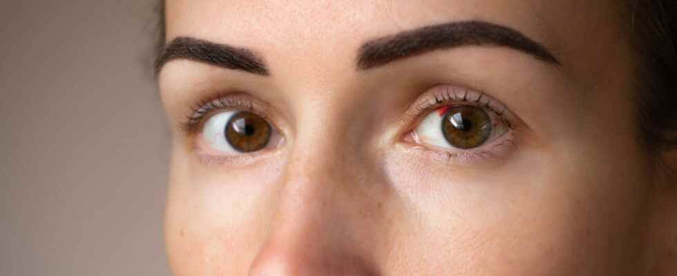
Retinal vein occlusion can involve the central retinal vein (CRVO) or a branch of the vein. An American study shows that the incidence of retinal vein occlusions seems to increase within 6 months of the diagnosis of COVID-19. A new symptom in sight?
[Mise à jour le 19 avril 2022 à 10h50] A new symptom of Covid or rather a consequence? According to an American study conducted on 432,515 patients positive for Covid-19 and published in the JAMA April 14, theincidence of retinal vein occlusions seems increase within 6 months following the Covid-19 diagnosis. More weakly for arterial occlusions of the vein (rarer and more serious). Covid can lead to systemic vascular damage, the researchers remind. Their cohort study included patients with no history of retinal vascular occlusion who were diagnosed with Covid infection between January 20, 2020 and May 31, 2021.maximum incidence venous occlusions and arterial occlusions occurred 6 to 8 weeks and 10 to 12 weeks respectively, after diagnosis of Covid. “These events remain rare, temper the authors in their conclusions, and in the absence of randomized controls, a causal relationship cannot be established.” For them, further large-scale epidemiological studies are warranted to better define the association between retinal thromboembolic events and Covid-19 infection. What’s this retinal vein occlusion? What causes usually? What to do ? What treatments ? Explanations with our doctor.
The retina, the nerve tissue lining the back of the eye that allows vision, is composed arteries and veins that bring oxygen and nutrients. This organ may suffer from vein occlusion which results in a stoppage of blood flow in the veins of the retina. “This phenomenon usually appears quite abruptly, and concerns mainly people over the age of 60, with associated cardiovascular risk factors and whose eye strain, which should normally be between 10 and 21mm of mercuryturns out to be too important, explains Dr Pierre Quéromès, ophthalmologist at the Quinze-Vingts National Ophthalmology Hospital Center and OPH 78. Vein occlusion is usually unilateralIt is very rare for both eyes to be affected at the same time. There are two types of occlusions:central retinal vein occlusion where the entire retina is affected, and the vein branch occlusions where only part of the retina is affected”.
Central retinal vein occlusion is primarily manifested by sudden drop in vision, distorted lines, blurred vision. On the other hand, she is painless and does not cause no redness, the eye is white.
Most commonly, central retinal vein occlusion is related to an obstruction of the vein by a small thrombus. “Some occlusions may also appear under venous spasmsas well as in the context of pathologies that are due to blood clotting. In these cases, vein occlusions may be bilateral and affect younger populations. These are atypical, rarer occlusions, which should lead to a blood test to look for other associated diseases such as cardiovascular pathologies (diabetes, cholesterol, alcohol, tobacco, obesity), ocular hypertension, glaucoma or even sleep apnea”says the eye surgeon.
CRVO occurs rarely in young people. It should lead to search for coagulation disorders.
Sudden loss of vision, distorted lines, black veil before the eyes, a painless white eye that is not accompanied by a dirty secretion or a burning sensation should to consult an ophthalmologist urgently. The doctor will be able to assess whether it is a vein occlusion or another pathology of the retina.
The diagnosis of CRVO is placed in consultation with an ophthalmologist. This will carry out various examinations:
- L’visual acuity to assess impairment of visual acuity;
- The eye pressure measurement because some serious vein occlusions can lead to neovascular glaucoma;
- A fundus which consists of placing drops in the eye to dilate the pupil and observe the retina.
“Depending on the signs observed at the back of the eye, the ophthalmologist will confirm or not the diagnosis of CRVO. In case of confirmation, a photo of the retina (retinophoto) or a retinal OCT will be carried out to evaluate the damage in the retina caused by vein occlusion: atrophy, retinal edema. An angiography (photo of the retina with injection of contrast product) can also be used to analyze the situation”says Dr. Pierre Quéromès.
“In a minority of people, there is a fairly rapid reperfusion of the vein and very little or no loss of vision, in which case it simply requires setting up fundus monitoring and performing optical coherence tomography (OCT), an imaging technique that allows exploration of the retina and cornea “, notes the eye surgeon. In the majority of cases, when the occlusion affects the entire retina or the macula, the treatment is based on intravitreal injections (IVT), sometimes associated with sessions of laser focal. Then, to protect the retina and avoid bleeding or neovascular glaucoma, laser by PPR in the periphery of the retina is indicated.
The prognosis is depending on the type of OVCR. In central retinal vein occlusions where the entire retina is affected, the visual prognosis for the future is poor, even with treatment. In branch vein occlusions, when only part of the retina is affected, the prognosis is better.
Thanks to Dr Pierre Quéromès, ophthalmologist at the National Hospital Center of Ophthalmology of Quinze-Vingts and at the OPH 78.
