Published on
Updated
Reading 2 mins.
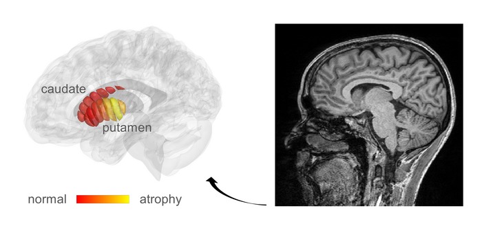
Researchers from the Hebrew University of Jerusalem have used a new imaging technique to diagnose Parkinson’s disease in its early stages. A real hope to change the diagnosis of patients but also to move towards personalized care.
Parkinson’s disease is a degenerative pathology, which affects dopaminergic neurons, in particular located within a particular cerebral structure, the striatum. Usually, its diagnosis is based on a clinical examination sometimes coupled with the realization of a cerebral MRI. But the sensitivity of this device does not reveal the biological changes that occur in the brains of Parkinson’s patients. It mainly helps to rule out other possible diagnoses. The signs of the disease are therefore only visible on this type of image at an advanced stage.
Images made using quantitative MRI
According to Israeli researchers, cellular changes linked to Parkinson’s disease could be visible by adapting a related technique, known as quantitative MRI (qMRI). By working on quantitative MRI images, they were able to examine the microstructures of the deep part of the brain, known as the striatum, which deteriorates during the onset and progression of Parkinson’s disease.
Identical images taken in different ways
Quantitative MRI images are obtained using different excitation energies. It is therefore the same structure examined each time, but under different conditions (a bit like taking the same photograph but with different lighting). Result: this technique made it possible to observe the initial changes within the examined brain tissue.

“When you don’t have measurements, you don’t know what’s normal and what’s abnormal brain structure, and what changes as the disease progresses.” write the researchers. “This new information will facilitate early diagnosis of the disease and provide “markers” to monitor the effectiveness of future drug therapies.”.
Such a degree of detail was previously only possible in specialized laboratories and on brain cells of post-mortem patients. Not ideal for early diagnosis or for monitoring the effectiveness of a drug!
Towards a personalized treatment for Parkinson’s disease
According to the authors, their tool can be set up and used routinely.in the next 3 to 5 years”. For Dr. Christophe De Jaeger, physiologist and specialist in aging, “when it is implemented in a few years, this technique could be used by clinicians to determine the presence or absence of Parkinson’s disease, in the most moderate cases where it is sometimes difficult to make a diagnosis. It is a less invasive method than what currently exists, such as brain scans which require the injection of radioactive product” first explains the doctor. “This could provide a strong diagnostic element allowing early management..
According to the authors, this discovery goes beyond simple diagnosis. According to them, it will make it possible to promote the search for new drugs and to identify subgroups of patients who may react differently to certain drugs. They see in this analysis “the first step towards a personalized treatment of Parkinson’s disease.
