
Published on
Updated
Reading 2 min.
A team of Swiss researchers have designed a brand new MRI procedure that can detect multiple sclerosis earlier and more accurately. An improvement for diagnosis but also for monitoring and understanding the disease.
Detecting multiple sclerosis (or MS) as early as possible becomes possible following the publication of a team of Swiss researchers. This team from the Swiss Federal Institute of Technology in Zurich has developed a new magnetic resonance imaging (MRI) procedure capable of assessing the severity and progression of autoimmune disease, with new reliability and precision.
An MRI capable of mapping myelin sheaths
How does MS work? As part of this autoimmune disease, the patient’s immune system begins to attack and destroy the myelin sheaths of the central nervous system. Similar to the insulating sheath that wraps an electrical wire, the myelin sheath allows the conduction of nerve signals (electrical impulses) along the nerve fiber. Unfortunately, when these sheaths are too damaged or too thin, movement, language and vision disorders can appear.
Until now, it was not possible to clearly visualize the myelin sheaths. This is because most of these devices work by reacting to water molecules in the body that have been stimulated by radio waves in a strong magnetic field. But the myelin sheaths, which surround nerve fibers in several layers, are mainly made of adipose tissue and proteins.

Unlike standard MRIs which construct their images primarily using signals from hydrogen atoms present in myelin water (content between two layers), the new MRI method ends this limitation by directly measuring the amount of myelin present in different areas of the brain, and by affixing numerical values to the imaging results. To do this, MRI must be capable of capturing much briefer signals from the hydrogen atoms contained in the tissue (of the order of a few microseconds) and providing a stronger magnetic field gradient. A challenge taken up by researchers who have developed a particular brain scanner with the companies Philips and Futura.
An evolution that can be discovered visually
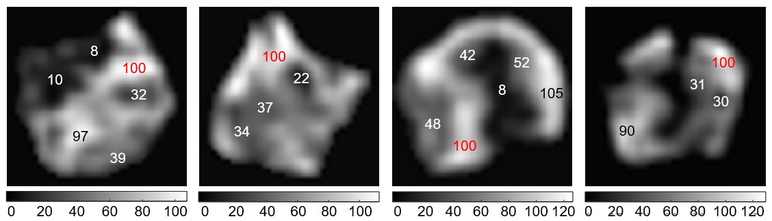
“The darker the area and the smaller the number in the image, the more the ducts have been reduced” indicates the press release which accompanies the publication. “The number 8, for example, means that the myelin content at this stage is only 8% of a maximum value of 100, indicating significant thinning of the myelin sheaths.” Of This way, it would become visually simpler to detect not only the possibility of MS, but also the progression of the disease.
The researchers have already successfully tested their MRI procedure on tissue samples from MS patients and two healthy people. They now plan to test it on patients affected by the autoimmune disease.
Remember that there is currently no cure for multiple sclerosis, only therapies aimed at reducing the inflammatory reaction. Recognizing the disease as early as possible would still make it possible to better care for each patient, slow down the progression of the disease and better support them.