Published on
Updated
Reading 3 mins.
Affecting millions of people around the world and known for centuries, migraine has not revealed all its secrets. A new study, presented at the annual meeting of the Radiological Society of North America (RSNA), reveals a change in certain structures of the brain of migraine sufferers.
Not comparable to a simple headache, migraine can be debilitating and prevent those who suffer from it from living normally. Seizures can cause nausea, vomiting, generalized weakness, and hypersensitivity to light. Worldwide, patient associations count more than 148 million migraine sufferers.
Visualize brain activity with ultra-high resolution MRI
If the symptoms of migraine are well described, what is happening in the brain is still quite mysterious. Scientists know the mechanism that causes pain, the dilation of vessels, and are beginning to discover effective therapeutic leads.
But the mechanism at the origin of these crises is not completely elucidated. To find out more, the scientists recruited 10 people with chronic migraine, 10 people with episodic migraine without aura, and five age-matched healthy controls. All patients were between 25 and 60 years old2.
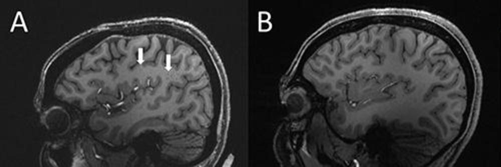
Ultra-high-field 7T MRI was used to compare structural microvascular changes in different types of migraine. Why ? “Because 7T MRI is able to create images of the brain with much higher resolution and better quality than other types of MRIs, it can be used to demonstrate much finer changes that occur in brain tissue. after a migraine “explains Wilson Xu of the University of Southern California in Los Angeles.
What are the perivascular spaces?
Xu and his colleagues sought to determine the association between migraine and enlarged perivascular spaces on images produced by MRI. The perivascular spaces are fluid-filled spaces surrounding the blood vessels of the brain.
Enlarged perivascular spaces may be a sign of underlying small vessel disease. “They are part of a fluid clearance system in the brain“, he added. “Studying how they contribute to migraine could help us better understand the origin of migraines.”
Enlarged perivascular spaces in the brain of migraine sufferers
Thanks to these 7T MRI images, the researchers were able to identify cerebral microhemorrhages. Their analysis further demonstrated that:
- The number of enlarged perivascular spaces in a region of the brain called the centrum semiovale was significantly higher in patients with migraine compared to healthy controls;
- The amount of enlarged perivascular space in the centrum semiovale was correlated with the severity of deep white matter hyperintensity in migraine patients.
Researchers go further and hypothesize that “significant differences in perivascular spaces in patients with migraine compared to healthy controls may suggest glymphatic disturbance in the brain“. The glymphatic system is a waste disposal system of the central nervous system.
Additional studies are still needed
“We studied chronic migraine and episodic migraine without aura and found that for both types of migraine the perivascular spaces were larger in the centrum semiovale (central area of white matter)“write the scientists.
“Although we found no significant change in the severity of white matter lesions in patients with and without migraine, these lesions were significantly related to the presence of enlarged perivascular spaces. This suggests that they may contribute to the development of these lesions“. A hypothesis that remains to be confirmed.
The researchers also cannot say whether these changes affect the development of migraine or are a result of the migraine…Scientists hope to be able to answer these questions through larger studies involving more diverse groups of migraine patients .
The opinion of Dr Wilfrid Casseron, neurologist
Asked about this work, Dr. Wilfrid Casseron, neurologist in Aix-en-Provence, is surprised first of all by the low number of participants in this study: “The results put forward by these researchers are interesting, but they do not highlight a clear link between these modified brain structures and migraine. But beyond that, it is the small number of participants recruited that is surprising: only 25 people in total is far too few to draw any conclusion from this study. It’s a shame, with the number of people who suffer from migraine, it would have been necessary to constitute a larger cohort“.