There is no shortage of superlatives. The most powerful MRI in the world has delivered, near Paris, its first images of the human brain with astonishing resolution. A step forward for scientists towards a better understanding of its functioning and of certain neurodegenerative or psychiatric diseases.
In 2021, researchers from the CEA, located on the Saclay plateau (Essonne), chose to debut the machine with a pumpkin, before the health authorities recently gave the green light for the examination of human subjects. It was necessary to prove to the health authorities that the intensity of the magnetic field has no effect on health.
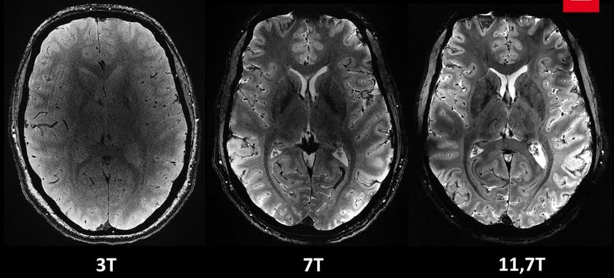
Over the past few months, around twenty healthy volunteers were able to enter the machine’s lair, which revealed the first images of their brain. “We have a level of finesse never before reached at the CEA,” says Alexandre Vignaud, physicist and research director at the CEA. This is the revolution: the magnetic field of this extraordinary magnet reaches 11.7 T (tesla), making it possible to obtain images ten times more precise than those currently produced in hospitals, where the power of MRIs does not exceed not 3 tesla.
Images from the Iseult MRI
© / CEA
On Alexandre Vignaud’s screen, images of brain sections are compared with what a 3 or 7 Tesla MRI would have given: “with this machine, we can see the very small vessels which supply the cerebral cortex, or details of the cerebellum which were almost invisible until now”, he comments. “Their precision is hardly believable!” enthused the Minister of Research Sylvie Retailleau in a statement to AFP. “This world first will make it possible to better detect and better treat brain pathologies.”
More than 20 years of research
The machine: a 132-ton magnet housed in a cylinder 5 meters long and as high, made up of a coil carrying a current of 1,500 amps, has an opening of 90 cm to accommodate a human body. This technical feat, the result of a Franco-German partnership, required more than 20 years of research. Named “Iseult”, the MRI (magnetic resonance imaging device) is the star of Neurospin, the CEA brain imaging research center, headed by neuroscientist Stanislas Dehaene. Two competing projects, in the United States and South Korea, have similar ambitions but have not yet reached the crucial stage of imaging on humans.
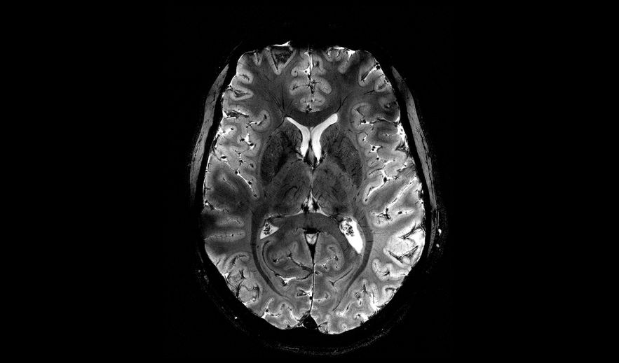
The first images of the brain with the Iseult MRI
© / CEA
One of the objectives of this extraordinary MRI is to refine the understanding of the anatomy of the brain and the areas which are activated when carrying out certain tasks. Scientists already know that different types of images that we are capable of recognizing (a face, a place, a word, etc.) activate distinct regions of the cerebral cortex. With MRI at 11.7 T, “we will be able to better understand the relationship between structure and cognitive functions of the brain, when we read a book or do a mental calculation for example”, assures Nicolas Boulant, director of research at the CEA and scientific manager of the project.
But it will also be a question of elucidating the mechanisms at work in neurodegenerative diseases (Parkinson or Alzheimer type) or in psychiatric conditions (depression, bipolarity, schizophrenia, etc.). “We know, for example, that a particular area – the hippocampus – is involved in Alzheimer’s disease, so we hope to be able to understand the organization and functioning of the cells in this part of the cerebral cortex”, illustrates Anne-Isabelle. Etienvre, director of fundamental research at the CEA.
The first images of the brain with the Iseult MRI
© / CEA
Researchers also hope to be able to map the distribution of certain drugs, such as lithium, used in the treatment of bipolar disorder. The very high magnetic field of the machine will indeed make it possible to identify the brain structures targeted by lithium in patients and to distinguish more or less good responders to treatment. “If we better understand these very impactful diseases, we should be able to make an earlier diagnosis, and therefore treat them better,” says Anne-Isabelle Etienvre.
Iseult will remain dedicated to fundamental research for a number of years. “The device is not intended to become a clinical diagnostic tool but we hope that the knowledge acquired can then be used in hospitals,” underlines Nicolas Boulant. In fact, the cost of this installation amounts to 215 million euros, too expensive to use it in a hospital setting. New healthy volunteers should be recruited by the end of summer. The brains of sick patients will not be studied for a few more years.