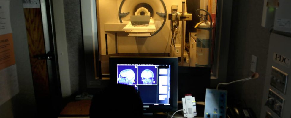
It’s a first. Images of our brain with great precision like we have never captured before. All this was possible thanks to the Iseult MRI, the most powerful MRI in the world. Around twenty volunteers passed through the running machine and were able to reveal previously unseen images of the brain. Report from the Saclay plateau of the Atomic Energy and Alternative Energies Commission (CEA), near Paris.
“ The Iseult magnet is the jewel of this installationexplains Alexandre Vignaud, research director at the Atomic Energy and Alternative Energies Commission (CEA). This magnet is five meters long, five meters in diameter, 132 tons. It is therefore there to generate the magnetic field which will be used to then obtain the MRI signal. “.
The magnet is the central element of the MRI, here it takes the form of a gigantic gray cylinder. The stronger the magnet in an MRI generates a magnetic field, the higher the quality of the images obtained. Iseult is the most powerful MRI machine in the world for taking images of humans. Its magnetic field is four to eight times more powerful than a hospital MRI, reaching 11.7 Tesla.
“ The Tesla is not a car, the Tesla is a unit of measurement of the magnetic field », specifies Alexandre Vignaud.
20 years of work
This machine is the fruit of 20 years of work. To achieve such power, the magnet materials must be in a state called “superconductor”. This is explained by Lionel Quettier, research engineer, who was project manager on the development of the magnet. “ To reach the superconducting state, we must cool materials, which we will cool to -271°C, therefore colder than your fridge. There are no commercial MRI magnets today that operate at this temperature. »
This level of intensity allows extremely fine parts of the brain to be observed. In front of the device’s on-board computer, Alexandre Vignaud marvels at the first images from the MRI. “ I zoomed in on the image and we actually realize that it is possible to see these tortuous ribbons which are in fact the cortex, that is to say where the neurons are. We can even see a contrast inside this ribbon, in particular small black streaks and a sort of equally black central channel. These are the blood vessels which supply the cortex, therefore which supply the neurons », adds the research director.
After the first tests, it’s time for science: this level of detail will allow us to better understand the brain and its diseases. It should help detect and treat certain pathologies, such as Alzheimer’s or Parkinson’s disease. New images of the brains of healthy patients will soon be made. The first images of diseased brains will not arrive before the end of 2025.
Also listenHow do smells affect our brain?
