Published on
Updated
Reading 3 mins.
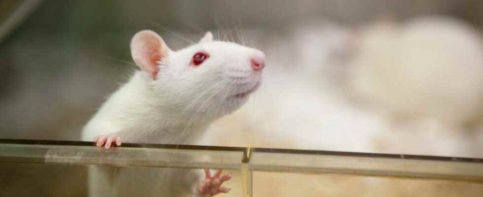
To better understand the development of human neurological disorders, such as schizophrenia, scientists transplanted human brain cells into the brains of young rats.
The information seems surprising, even disturbing, and yet: scientists have implanted organoids – human multicellular micro-tissues – in young rats to better study the occurrence of certain psychiatric disorders. The results of the study are available in the journal Nature.
Young rats were used
As part of this study, the scientists implanted brain organoids – tiny structures made from human stem cells – into the brains of young rats, and more precisely into a region called the cerebral somatosensory cortex.
The age of the rodents was essential because the brain of the rodents had to continue to develop, for the good integration of the human cells.
However, the experiment seems to have been conclusive, because by using these cells on a young animal, “we found that organoids can become quite large and vascularized“, and be perfectly irrigated to the point “to occupy about a third of the hemisphere of the brain of the animal”, reveals Sergiu Pasca, professor of psychiatry and behavioral sciences at the American University of Stanford (California), and main author of the study.
However, the researchers waited for the brains of the rats to mature (+6 months) before observing the perfect cerebral integration of the organoids.
This is exciting news for scientists, although this transplant procedure is probably still too expensive to become a real research tool.
A hope for better understanding and treating brain diseases
It is very difficult to study mental disorders because animals do not experience them in the same way as humans, who cannot be experienced in vivo.
Scientists are already practicing cultures, in Petri dishes, of human brain tissue from stem cells.
But in the laboratory, “lNeurons don’t grow to the size they would in a real human brain“, explains Sergiu Pasca, professor of psychiatry and behavioral sciences at the American University of Stanford, and main author of the study published in Nature.
Moreover, since these tissues are cultured outside the human body, they do not allow the symptoms resulting from a defect in their functioning to be studied.
The solution is to implant these human brain tissues, called organoids, into the brains of young rats. Age is important because the brain of an adult animal stops developing, which would have affected the integration of human cells.
By transplanting them to a young animal, “we have found that the organoids can become quite large and vascular” and therefore be supplied by the blood network of the rat, to the point of occupying about a third of the hemisphere of the brain“of the animal, details Pr. Pasca.
The researchers tested the correct implantation of the organoids by sending a blast of air to the rat’s whiskers, which resulted in electrical activity in the human-derived neurons – a sign that they were playing their role as receivers well with a stimulus.
They then wanted to know if these neurons could transmit a signal to the rat’s body. To do this, they implanted organoids previously modified in the laboratory to react to blue light. They then trained the rats to drink from a cannula of water when this blue light stimulated the organoids via a cable connected to their brains. The maneuver proved effective in two weeks.
Ethical issues
The team finally used their new technique with organoids from patients with a genetic disease, Timothy syndrome. She observed that in the brain of the rat, these organoids grew more slowly and had a lower activity than organoids from healthy patients.
This technique could eventually be used to test new drugs, according to two scientists who were not involved in the study, but who commented on its findings in Nature.
She “takes our ability to study human brain development, evolution and disease into uncharted territory“, write Gray Camp, of the Swiss Roche Institute for Translational Bioengineering, and Barbara Treutlein, of the Zurich Institute of Technology (ETH).
The technique raises ethical questions, in particular that of knowing to what extent the implantation of human brain tissue in an animal can change its deep nature.
Professor Pasca ruled out such a risk for the rat, because of the great rapidity with which its brain develops compared to that of a human. He described as a “natural barrier” the functioning of a rat cortex, which would not have time to deeply integrate neurons of human origin.
On the other hand, such a barrier could not exist in species closer to humans, according to Prof. Pasca, opposed to the use of this method in primates.
It underlines the moral imperative “to be able to better study and possibly treat psychiatric disorders, while taking into account the proximity to humans of the animal model used.
“Human psychiatric disorders are very largely unique to humans. That’s why we’re going to have to think very carefully (…) how far we want to work on some of these models“, according to him.
