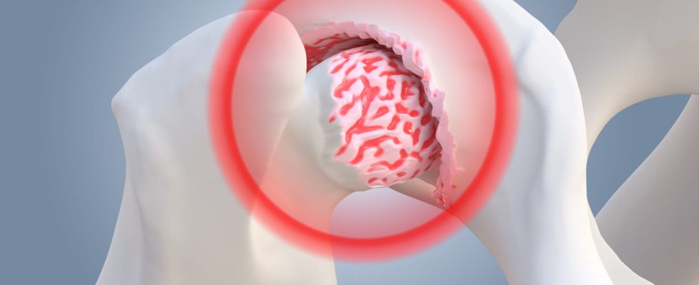
Hip dysplasia is a malformation of the femoral head embedded in the joint cavity of the pelvis. Its symptoms appear more generally in adulthood and young women are more affected.
What is hip dysplasia?
“The hip is a joint made up of two parts: the femoral head, like a “billiard ball”which is covered and embedded in the basin in a hemispherical cavity, the acetabulum. When the latter is not covering enough. The femoral head is exposed and this is called hip dysplasia.“, explains Dr Alexis Nogier, specialist hip surgeon in Paris. The definition of hip dysplasia is radiological : the angle of bone coverage will determine the degree of dysplasia.
What are the symptoms ?
Initially there are no symptoms: This is a malformation that is not painful. People with dysplasia generally have fairly wide hip mobility. The mechanical conditions of the hip in cases of dysplasia are not favorable and it is in adulthood that the symptoms appear :
- of the mechanical painparticularly when walking, located at the level of the gluteus medius or groin
- pain in prolonged static position
- pain while playing sports
- a limp
- cracking in the hip
Advice before treating dysplasia through surgery is given to the patient to eliminate the pain(s), in particular weight loss, “the hip supporting 2 to 3 times the weight of the body“, modulation of sporting activity: avoid sports with load and impact (running) and prefer cycling or swimming. Muscle strengthening of the gluteus medius will help: the more the gluteus medius will be muscular and sheathed, the less painful the hip will be.
When hip dysplasia is diagnosed as severe, surgical procedures can be proposed to correct it:
- Periarticular osteotomy which consists of cutting the pelvis to correct the dysplasia
- The bone stopa little rarer as an intervention: a piece of pelvic bone is screwed above the femoral head to improve its coverage
- When there is fairly severe wear of the cartilage, we suggest placing a hip prosthesis.
What causes hip dysplasia?
Hip dysplasia is of origin congenital : “it’s a hip at birth that is not well formed and which, as it grows, will not develop in a normal way: its joint will be flexible and stable but the bone coverage will be insufficient“, specifies our interlocutor. It is indeed a malformation of the bone structure of the hip.
What are the consequences for development?
In extreme forms of dysplasia, we can end up with congenital hip dislocation : the femoral head will come out of the acetabulum. “These are generally very specific cases diagnosed at birth. or in the first years of life, because there are difficulties acquiring walking or lameness“, notes Dr Nogier. For most hip dysplasias, there are no consequences on the child’s life. The first consequences arrive in adulthood when pain appears and allows the diagnosis to be made.
The diagnosis of hip dysplasia is radiological and will correspond to the measurement of the lateral femoral coverage angle. An x-ray of the pelvisstanding and facing, will make it possible to define this coverage angle which must normally be greater than 25°
- Between 20 and 25°dysplasia is limit
- Between 10 and 20°dysplasia is effective
- Less than 10°dysplasia is severe.
Next, we will observe whether hip dysplasia is not associated with other malformations at the level of the roof of the acetabulum or the femoral head. In consultation, we will also be interested to lesions associated with hip dysplasia. “In a rather young woman who suffers from pain in the groin or trochanter, we will suggest an MRI or arthroscopy looking for lesions associated with his hip dysplasia: lesions of the labrum, lesions of the cartilage (chondropathy), damage to the subchondral bone and signs of osteoarthritis“, he lists.
Thanks to Dr Alexis Nogier, hip surgeon, Paris
