You will also be interested
[EN VIDÉO] 3D brain produced using 3D MRI Using the new IRMa 3D software, a 3D animation can be created from an MRI image. The vivid detail of these animated amplified movements can help identify abnormalities, such as those caused by blockages of spinal fluids, including blood and cerebrospinal fluid in the brain.
The Covid-19 still occupies researchers, who are very interested in the long-term consequences of mild to moderate forms of the disease. In an article published on March 7, 2022 in Naturea research group from the University of Oxford, England, compared “before-after” brain images of infection from several hundred patients with those from uninfected people.
Brain abnormalities even in moderate forms of Covid-19
English scientists have analyzed 785 scanners brain of theUK Biobankthe database the largest medical procedure in the country, carried out on people aged 50 to 80. Of these patients, 401 tested positive for SARS-CoV-2 between the first examination and the second, which takes place on average 141 days after the diagnostic of Covid-19, and the others were not infected. For the comparison of the pictures to be relevant, the scientists made sure that they had been obtained by the same methodology and with the same device at MRI Everytime.
The images taken before the infection make it possible to ensure that the anomalies visible are not due to a pre-existing disease. Infected patients show a reduction in the thickness of the matter gray in the prefrontal cortex and the gyrus parahippocampus, significant tissue damage in regions whose function is related to the olfactory cortex, and an overall reduction in brain size. What’s more, cognition of infected patients also declined between the two scanners.
Surprising results since the patients included only developed a mild to moderate form of Covid-19. They remain significant even when the 15 hospitalized patients are removed from the analysis. These abnormalities could be the trace in the brain, left by the proliferation of SARS-CoV-2 in the olfactory bulb and the resulting anosmia, or inflammatory events observed in some patients. A long-term follow-up will make it possible to know if these anomalies are reversible or persistent.
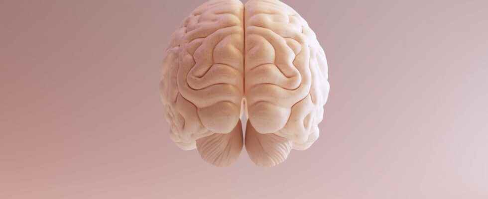
The damage of Covid-19 on the human brain in pictures
Article published on December 12, 2021 by Julie Kern
Covid-19 attacks the respiratory tract but also the brain. During its annual congress, the Society of radiology of North America presented the results of its study on theimpact brain damage in Covid-19 patients. About 1% of the patients followed are affected by various abnormalities visible after an imaging examination.
Covid-19 is a disease that affects many organs. If respiratory problems are the most emblematic and the most studied, the brain is not spared. The Radiological Society of North America (RSNA for Radiological Society of North America, in English) wanted to determine the incidence of brain damage in patients with confirmed SARS-CoV-2 infection. ” Our study shows that complications in the central nervous system represent a significant cause of morbidity and mortality in this pandemic devastating said Thomas Jefferson, director of the radioneurology unit at the University of Philadelphia. Of the 39,750 patients cared for in the 11 medical centers that participated in the study (in the United States and Europe), 4,342 underwent a medical brain imaging examination. And for 10% of them, the images showed visible abnormalities.
Brain damage in people with Covid-19
Patients are on average 65.8 years old and men are twice as numerous as women. The most frequent complication is thestroke for 62% of patients, followed by intracerebral hemorrhage andencephalitis. Most often, these lesions are unilateral, that is to say located on only one side of the brain. The regions most affected are the frontal and parietal lobe in more than one out of two cases, followed by the lobe temporalfrom cerebellum and finally of brainstem. The lesions are as much located in the white matter, cortical or subcortical.
And the developing brain?
During this same congress, the RSNA also studied the effects of Covid-19 on the brains of babies. A mild to moderate infection during the pregnancyaround the 28and week in the case of the women studied here, does not appear to alter the brain development of the fetus. ” In our study, there was no evidence that maternal SARS-CoV-2 infection has any effect on the brain development of the unborn child. explains Dr. Sophia Stöcklein of the Ludwig Maximilian University of Munich. In addition, proven cases of vertical mother-to-child transmission of SARS-CoV-2 are very rare and their effect on fetal health remains to be determined.
Interested in what you just read?