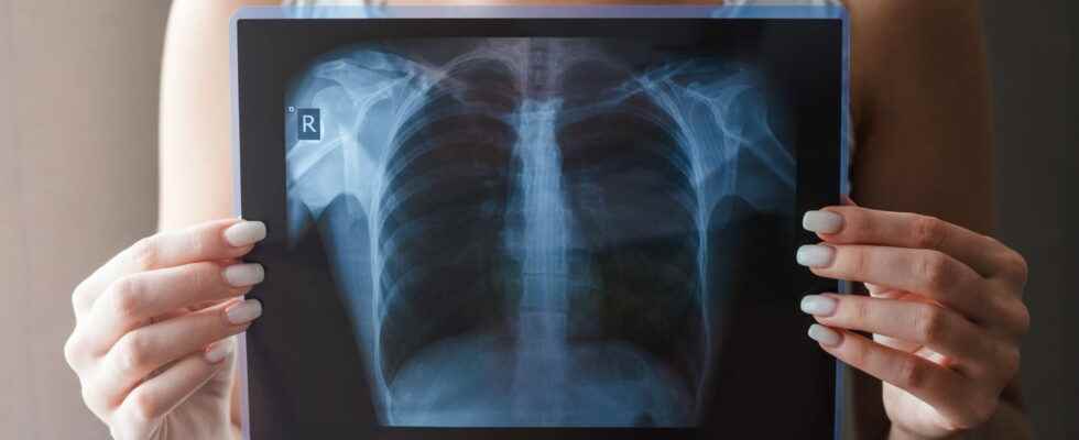
Indicated to detect abnormalities or pulmonary or arterial pathologies in the rib cage, the chest CT scan is a relatively frequent, non-invasive and painless examination of the lungs. Do you have to be fasting to pass it? When do we get the results? What are the risks ?
Like radiology, the scanner uses x-rays, thanks to a tube/detector pair rotating in “continuous rotation” around the patient. The images are then processed and reconstructed to give images of the explored structures. It’s about a 3D volume image reprocessed “in sections” in the 3 planes (even obliquely). The chest examination takes only a few seconds, several hundred sections of 1 millimeter can be obtained, allowing a very fine analysis. It’s a simple, fast and non-invasive examination. One iodine-based contrast agent can be injected intravenously in order to clearly differentiate the vessels from other tissues, or to reveal abnormal structures. The organs studied with a chest scanner are:
- lungs,
- the mediastinum: the central axis of the rib cage, located between the two lungs, which connects the neck and the abdomen, containing among other things,
- thyroid,
- Ships,
- the heart,
- the esophagus,
- the trachea
- the two main bronchi.
A chest CT scan can be done when:
- An infection such as pneumonia, bronchitis, pneumonia
- Diseases of the supporting tissue of the lung: fibrosis, sarcoidosis…
- Emphysema (when air gets “stuck” in the lung)
- dilation of the bronchi
- Exposure to asbestos or other pollutants
- Pleural pathologies, in particular mesothelioma
- Screening (protocols being validated), diagnosis and extension assessment of all malignant or benign thoracic tumours. the study of possible metastases in the lungs.
- Search for a pulmonary embolism or dissection of the aorta, study of the coronary arteries.
- A trauma assessment: rib fracture, thoracic contusion, blood effusion (hemothorax), vascular lesion…
The patient is lying down with his arms behind his head.in order to avoid annoying bone artifacts, on a bed that moves in a wide ringmost of the time on the back (you may have to make an acquisition on your stomach in rare cases). The patient is alone in the examination room, the medical team is close to him, behind glass. She sees and hears him throughout the examination and communicates with him via a microphone and can thus intervene at any time if necessary. The exam is usually quick (less than 10 minutes). The cooperation of the patient is essential: he must try to stay stillit will be specified to him when he must refrain from breathing for a few seconds. If the examination requires an ‘injection of contrast product’, this is done by intravenously during the examination, using an automatic injector. The only pain likely to be felt is that of the bite, which is light and transient. “But, it is not uncommon to feel a sensation of warmth at the time of the injection, an urge or feeling to urinate, or an odd taste in your mouth. With the latest contrast products, these sensations are less and less present.” warns Dr. Franck Clarot, radiologist.
“There are injection of iodine (contrast product) in the case of vascular pathologies, when, for example, a pulmonary embolism is suspected or when a check-up of the coronary arteries (coroscanner) is desired; and in the cancerous pathologies, in order to visualize masses, lymph nodes, and tumors“explains Dr. Clarot. For other indications, the thoracic scanner is generally done without injection. “Nevertheless, the radiologist can decide on an injection in certain cases in order to facilitate the reading of doubtful images..” says the radiologist.
The scanner images are available immediately after the exam but the radiologist must absolutely analyze them in their entirety, section by section, in order to be able to interpret the examination. It may take a while. The results are written in the form of a report, given to the patient or sent to the doctor applicant for the examination, accompanied by a CD or DVD containing the images of the examination. “Some so-called “key” images can also be reproduced on film or on paper; The exam can then be reread either on a dedicated console, or using a light viewer generally supplied on the CD/DVD medium. “explains Dr. Clarot.
It is not necessary to be fasting for a chest scan. However, it is preferable to eat light at the previous meal when the scan is done with injection: this prevent possible nausea. On the other hand, it will then be necessary to drink well in order to evacuate the product. The patient should also refrain from smoking before the examination. In all cases, unless otherwise advised by the attending physician or radiologist: you should stay hydrated before and after a CT scan (at least 1.5 liters of water per day).
- In case of kidney failure, even moderate: it is imperative to report it to the radiologist, who may decide not to perform an iodine injection. A recent blood test (less than 3 months) including creatinine clearance is systematically requested after 60 years.
- In case of diabetes treated with Metformin tablets (Glucophage®, Glucinan®, Stagid® and generics), the medication must be interrupted during the 2 days following the scan, and resumed after the creatinine assay because there is a risk of lactic acidosis
- In case of pregnancy or risk of pregnancythe examination will be postponed, except in a life-threatening emergency
The chest CT scan itself does not cause any side effects. On the other hand, the injection of the contrast product can lead to hot flushes, nausea, hives, and rarely vagal discomfort. Exceptionally, a sudden drop in blood pressure may be observed, thus requiring adequate therapy.
A chest scanner is listed ZBQK001 (or ZBQH001) in the SS nomenclature, for a cost of 25,27€ (which corresponds to the intellectual act of interpretation), to which is added a technical package corresponding to the equipment (of variable value depending on the geographical area, the class of equipment, the number of examinations carried out, etc.). The technical package is 100% supported, and it is often “transparent” and unknown to the patient (between €50 and €90 in general) Reimbursement varies according to several criteria: 100% coverage or not, CMU, general plan, course of treatment or not, etc. To these sums may be added a possible excess fees.
Thanks to Dr. Franck Clarot, radiologist. Statements collected in 2020.