MIT researchers have developed self-adhesive patches that allow organ ultrasounds to be performed for 48 hours. A small revolution that would be useful to control the evolution of a tumor, a pregnancy, or to avoid the consequences of overtraining for top athletes.
You will also be interested
[EN VIDÉO] Video: how an ultrasound works Ultrasound allows us to visualize the inside of the human body in a non-invasive way. It uses ultrasound to achieve this. Unisciel and the University of Lille 1 tell us more during this episode of Kézako.
Do a ultrasound in high definition by applying the equivalent of a small self-adhesive bandage to the skin, this is what researchers from the famous Massachusetts Institute of Technology (MIT) in the United States. Rather than having a lot of equipment and monopolizing an operator to drag and move a probe with freeze at the targeted body location, this patch can be worn by the patient for 48 hours and capture images of organ function during many activities. The operation of the patch is equivalent to that of a probe. It therefore issues ultrasound to represent the patient’s organs in imagery.
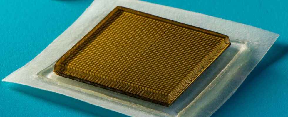
For the time being, this process remains experimental, but the researchers were able to observe the effectiveness of this patch for taking live, high-definition images of the main blood vessels and deeper organs such as the heart, lungs and thestomach. Specifically, the self-adhesive patch measures approximately 2 square centimeters in diameter and 3 millimeters thick. Between two layers of adhesive and stretchable elastomer that stick directly to the skin, there is a hydro gel solid that facilitates the transmission of ultrasound, since it never dries out. The assembly supports a rigid array of transducers, which helps generate clearer and more accurate images according to the researchers.
The hydrogel never dries out and it is this that ensures good definition of organ imaging. © MIT
A postage stamp-sized ultrasound machine
With its adhesive part, the patch can remain in place for at least 48 hours and, thanks to its wireless transmission, rather than picking up a situation punctually, it is able to collect changes in the organs while the patient performs different activities. .
During the tests, ultrasound was performed while the volunteers stood, sat, or ran or cycled. The deformation of the organs or the impact of a position or an activity could thus be examined. This is how the researchers were able to see in real time the temporary muscle microlesions that could be created when a volunteer lifted weights.
The researchers were able to see in real time the temporary muscle micro-lesions that could be created when a volunteer lifted weights
Scientists are already convinced of the immediate interest of this invention. It could already make it possible to carry out permanent monitoring of a patient’s organs, without having to immobilize or hospitalize him, for example. These patches could very well be purchased directly from a pharmacy.
Going a little further, by connecting the patch via Bluetooth to the smart phonea application dedicated could operate a AI decentralized to directly analyze the collected imagery. The follow-up of a tumor or a pregnancy could then be achieved by affixing this patch and making ultrasounds regular.
This project is not the only one around the miniaturization of ultrasound imaging. Further experimentation with portable and flexible systems for probing organs have already been tried, but the results remained mediocre concerning the definition. The devices were stretched to follow the roughness of the body and the image distorted accordingly. Furthermore, it was not possible to perform deep organ ultrasounds. Today, with this ultrasound patch, these obstacles seem to have been removed.
Interested in what you just read?