Published on
Updated
Reading 2 mins.
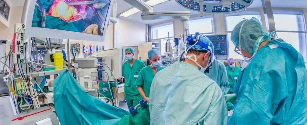
During surgery, it is very common for the surgeon to send an anatomical anatomo-pathology piece, so that they can find out more about the nature of the tumor that they need to remove. To speed up this waiting time, researchers have developed a new method based on artificial intelligence.
When operating on a patient, whether for a tumor or otherwise, the amount of tissue to be removed is usually not planned in advance. Different characteristics help the operator to decide: the nature of the tumour, its stage or even its location.
The anatomopathological analysis is quite long
Elements which are added to the analysis carried out by the anatomo-pathologist on a tissue sample, which he receives and analyzes during the operation or after, within 12 to 48 hours.
In the event that the analysis is immediate, this stage is shorter, the surgeon waits for a few minutes for the result, while the patient is on the table. The pathologist then uses a method to freeze the tissues, cut sections to observe these thin slices under the microscope.
Cryosectioning takes minutes, but can distort cellular details, compromise or tear delicate tissue. In addition, it has the disadvantage of creating artefacts which can complicate the diagnosis.
Artificial intelligence could help surgeons
According to the authors of this work, attached to the Mahmood Lab and the Brigham and Women’s Hospital in Boston, United States, a method based on artificial intelligence could be the solution.
AI would be a way to improve the quality of images to increase the accuracy of diagnoses, which would then be delivered more quickly. The authors demonstrated that the method could be used to “subtype” different types of cancer, including glioma and non-small cell lung cancer. Their results are published in the journal Nature Biomedial Engineering.
A method validated by pathologists
The team validated their findings by recruiting pathologists to make a diagnosis from images with AI and traditional cryosection images. The artificial intelligence method not only improved the image quality, but also increased the diagnostic accuracy of doctors.
For one of the authors, Prof. Faisal Mahmood of the Division of Computational Pathology at Brigham and Women’s Hospital, “Establishing a rapid diagnosis from frozen tissue samples is difficult and requires specialized training, but this type of diagnosis is a critical step in the management of patients during surgery.”
Next step: conduct clinical studies
The scientists believe that now their work must move to the prospective clinical study stage in order to validate the AI method and determine its use in real conditions. “Our work shows that AI has the potential to make critical, time-sensitive diagnosis easier and more accessible to pathologists.” adds Professor Mahmood. “And this could potentially be applied to any type of cancer surgery. It opens up many possibilities for improving diagnosis and patient management.”