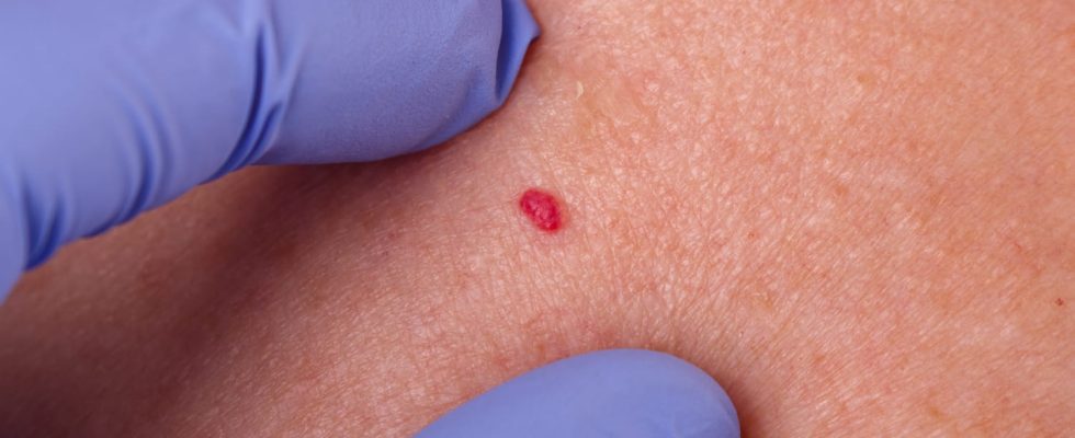
Stellar, cavernous or flat, the angioma looks like a spot on the skin. It can disappear spontaneously or last a lifetime.
Sometimes also called “superficial vascular abnormalities”, angiomas are defined by malformations located in the blood vessels, veins, arteries or capillaries or lymphatics. Most angiomas appear at birth, but others are acquired later. There are several forms of it, which are manifested by spots on the skin, of different color, size and shape, on the face, trunk, arms, legs. They usually end up fading or disappearing spontaneously, but some spots can also develop in adulthood. Although the vast majority are benign and develop on the skin, some angiomas can also be found inside the organs, which can sometimes lead to complications.
An angioma is a malformation caused by an exaggerated dilation of blood or lymphatic vessels. In the first case, we will speak ofhemangiomasin the second of lymphangiomas. Concretely, this malformation results from malformed excess vessels which seem to be the result of disorders occurring during their formation in utero.
These spots do not hurt.
These spots generally cause no pain and have, for the most banal forms, no clinical impact.. In some rarer cases, they can ulcerate and become painful. It is also possible that haemorrhages appear spontaneously (by necrosis) or after a local traumatism (in particular when they relate to the organs).
There are several kinds of angiomas:
- cavernous angiomas (cavernoma),
- spider angiomas,
- flat angiomaswhich is often called “birth mark” because they are present from birth and do not disappear spontaneously.
It should not be confused with infant hemangiomas which appear soon after birth, increase in size over the first 6 to 12 months of life, but often fade before adolescence.
In the majority of cases, angiomas appearing at birth are benign. However, it is essential to have them monitored by a pediatrician or dermatologist.
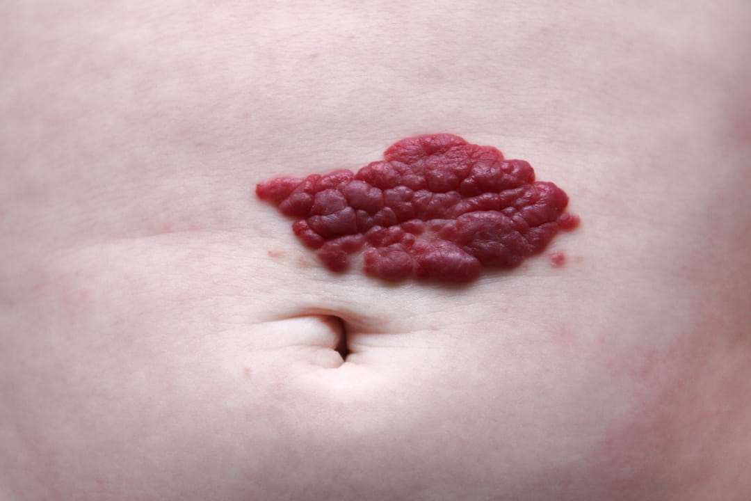

Angiomas such as “birthmarks” or “port wine stains” can be seen with the naked eye, as well as those that are not present at birth but disappear afterwards. The angiomas present on the organs generally discovered fortuitously during a CT scan, ultrasound or MRI. In this case, and depending on the affected organ, a medical specialist will have to confirm that it is indeed an angioma. When an angioma is discovered, it is nevertheless advised to carry out a medical follow-up, in order to prevent possible and very rare complications.
The treatment of the angioma depends on its type and location. Some drugs like beta-blockers or topical corticosteroids can be prescribed. Depending on his functional or aesthetic disability, surgical treatment is possible: excision, coagulation with an electric bistoury, laser (indicated for unsightly flat angioma), radiotherapy or radium, cryotherapy or embolization (the vessel in question is artificially blocked). But in the vast majority of cases, no treatment is put in place because they resolve on their own. However, they should be checked regularly. Local care is necessary in case of ulcerated lesions to avoid bleeding and infection. Laser treatment allow the targeted destruction of “abnormal” blood vessels in number and size which constitute the angioma.
There is no way to prevent angioma of any form. Angiomas are due to an unpredictable vascular abnormality. Upon discovery of an angioma, it is nevertheless advisable to carry out a medical follow-up, in order to prevent possible and very rare complications.