
Published on
Updated
Reading 2 mins.
Researchers have discovered the presence of specific markers in the brains of children with attention deficit disorder with or without hyperactivity (ADHD). This discovery could make diagnosis more efficient and reliable.
According to the High Authority of Saté, 3.5 to 5.6% of school children suffer from attention deficit disorder with or without hyperactivity (ADHD) in France, a disorder characterized by difficulty concentrating, impulsivity and agitation. In an attempt to improve the diagnosis and management of this disorder, researchers at Yale Medical University examined hundreds of children.
Abnormal connectivity in brain networks
The researchers used MRI data from the Adolescent Brain Cognitive Development (ABCD) study, the largest long-term study of brain development and health in children in the United States. The ABCD study involves 11,878 children between the ages of 9 and 10 at 21 centers across the country. After adjustments, Prof. Huang Lin’s study group relied on data from 7,805 patients, including 1,798 diagnosed with ADHD.
All underwent structural MRI, diffusion tensor imaging, and resting-state functional MRI. So many imaging exams to visualize brain activity and organization in these children.
The researchers then performed a statistical analysis of the imaging data to determine the association of ADHD with neuroimaging parameters, including brain volume, surface area, white matter integrity and functional connectivity.
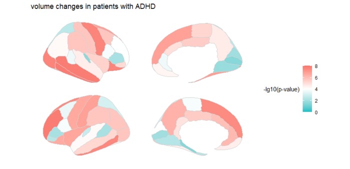
Result: In children with ADHD, scientists detected abnormal connectivity in brain networks related to memory and hearing processing, as well as thinning of the cerebral cortex and significant changes in white matter microstructure , particularly in the frontal lobe of the brain.
Multiple brain changes and not just an externalizing behavioral syndrome
“We found changes in almost every brain region we studied“, reveals Huang Lin, co-author of the study. “The pervasiveness throughout the brain was surprising, as many previous studies have only identified changes in specific regions.“.
“The frontal lobe is the area of the brain involved in managing impulsivity and attention or lack thereof – two of the main symptoms of ADHD”Lin said.
Be able to better diagnose ADHD tomorrow
According to the researchers, the MRI data are sufficiently important to be able to “feed” an algorithm capable of making a diagnosis of ADHD tomorrow. Machine learning, a type of artificial intelligence, is used to analyze large amounts of MRI data.
For the researcher, this step is crucial because the diagnosis of ADHD is still too unreliable today.
“Sometimes, when a clinical diagnosis is in doubt, objective brain MRIs can help clearly identify affected children.” says Huang Lin. “Objective MRI biomarkers can be used for decision making in ADHD diagnosis, treatment planning and treatment monitoring“.
The researchers’ findings will be presented during the 108th meeting of the Radiological Society of North America (RSNA), to be held in Chicago from November 27 to December 1, 2022.