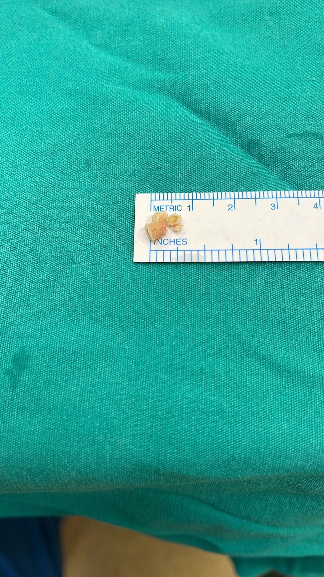
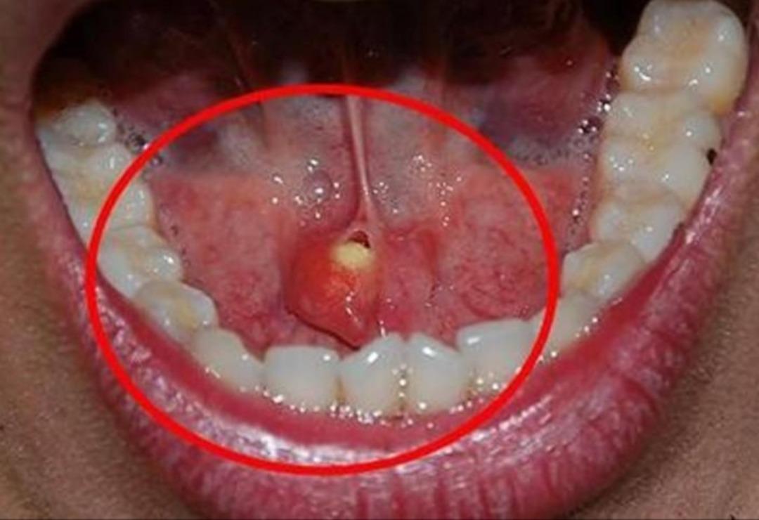
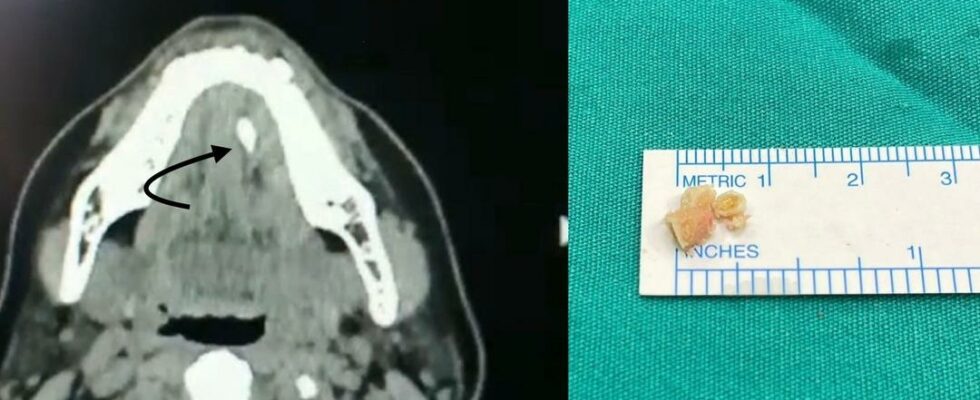
Stones formed in the salivary glands attract attention as an issue that is often ignored. However, experts say that these small stones can cause big problems. Stating that stones that accumulate in the salivary glands, medically known as sialoliths, can cause discomfort and pain in the mouth, ENT Specialist Assoc. Dr. Nesrettin Fatih Turgut talked about the diagnosis, treatment and post-treatment process of the disease.
BEWARE OF THESE SYMPTOMS
Salivary gland stones are stones seen in the salivary glands that produce saliva secretion, located under the chin and behind the cheeks, or in the salivary gland ducts that allow the salivary glands to open into the mouth, ENT Specialist Assoc. Dr. Nesrettin Fatih Turgut said, “Salivary gland stones are more common in men between the ages of 30-60, in the submandibular salivary glands and ducts under the jaw, because the density of saliva content is high. Insufficient fluid intake, infections that cause decreased salivary secretion, drug use and various infections, and stenosis of the salivary gland ducts predispose to the formation of stones in the salivary gland duct. The typical symptom of this disease is swelling and pain that develops after eating in the gland on the side where the stone is present. Accumulation of saliva secretion that cannot be expelled creates a predisposition to infection. A disease called bacterial salivary gland inflammation may develop, in which case the complaints may become severe. Excessive swelling, hypersensitivity, pain and fever of the affected salivary gland may develop. “If left untreated, we may progress to a condition that requires hospitalization and is more severe, deep neck infection,” he said.
“DEPENDING ON THE SIZE AND LOCATION OF THE STONE, SURGICAL INTERVENTION MAY BE AVAILABLE”

Assoc. Prof. stated that traditional treatments are recommended for patients whose complaints are milder and whose stones are small in size and located near the end of the salivary gland duct. Dr. Nesrettin Fatih Turgut said, “Painkillers are beneficial. Consuming plenty of fluids and applying heat can provide relief. We recommend consuming plenty of fluids to all our patients. At the same time, sucking sour products such as lemon increases salivary fluid and can help remove very small stones. Surgical procedures are considered in cases where the stone size is large and the stone is located close to the gland. The location and size of the stone and the condition of the affected salivary gland determine the type of surgery. If the stone is located in the salivary gland duct, localization and removal with a camera system called sialendoscopy is preferred without any incision. However, in cases where the stone is located in the salivary gland and its size is very large, surgical options with an external or intraoral approach come to the fore,” he said.
“LARGE STONES ARE REMOVED BY SHINING THEM USING THE AIR CRUSHING TECHNIQUE”

Talking about sialendoscopy, which is a method of examining the salivary ducts using a camera system that can reach the most extreme parts of the salivary gland ducts, Assoc. Dr. Turgut said, “This system provides a tool for the diagnosis and treatment of diseases within the canal. The main feature of the camera system is the ability to inspect the inside of the salivary duct to millimetric dimensions, a size of approximately 1.5 mm is mentioned. This procedure can be performed under general anesthesia or local anesthesia. Treatment planning is made depending on the physician’s experience, the patient’s health condition, the patient’s compliance and preference. Sialendoscopy is generally used in the treatment of patients with stones in the salivary ducts. This method can also be applied to patients with Sjögren’s disease, patients who have received radioactive iodine treatment, and pediatric patients with recurrent salivary gland inflammation. Sialendoscopy time may vary depending on the size and location of the stone. Large stones are removed by reducing them to size using the air crushing technique, so the processing time may be long. No incisions or stitches are applied during sialendoscopy, so no pain or complaints are observed after the procedure. There may be temporary swelling in the salivary gland on the same side, but this swelling usually subsides within 1-2 hours. “Patients are usually discharged on the same day,” he said.
“OPEN SURGERY CAN BE APPLIED FOR STONES THAT CANNOT BE REMOVED WITH SIALENDOSCOPY”

Turgut, who also gave information about the surgical intervention option, said, “It may not be possible to remove the stone with sialendoscopy due to reasons such as the large size of the stone, the stone being located in the gland, the stone being stuck to the canal due to frequent infection. In these cases, the option of open surgery comes to the fore. If the stone is located in the canal, the stone can be reached through a small incision made on the canal from inside the mouth and the stone is removed. The procedure is completed by placing a few stitches. In our patients, only if the stone is in the salivary gland or if the salivary gland has lost its function (atrophyed) due to continuous (chronic) infection, the salivary gland is completely removed by making an incision under the chin under general anesthesia. “Hospitalization may be required for 2-3 days after the surgery,” he said.
(UAV)
