Published on
Updated
Reading 2 min.
in collaboration with
Dr Gérald Kierzek (Medical Director)
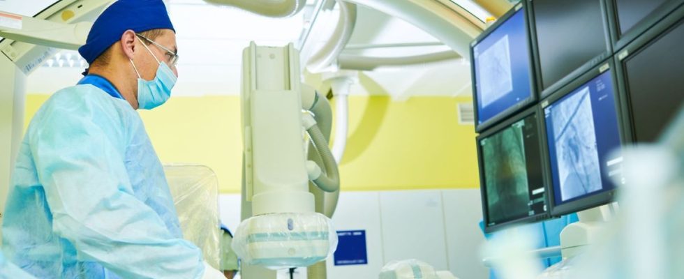
This is a world first: a Canadian neuroradiologist, Dr. Robert Fahed, has managed to film the inside of a cerebral blood vessel. He achieved this feat on a patient in his fifties, victim of repeated strokes, due to a malformation of the carotid artery, one of the arteries which supplies the brain with oxygen.
Canadian neuroradiologist, Dr. Robert Fahed, succeeded in filming the inside of a cerebral blood vessel using a camera attached to an optical fiber. This feat took place on November 14, 2023 at the Ottawa hospital, in Canada.
A camera designed specifically for cerebral arteries
The Canadian patient chosen, aged around fifty, was chosen because of a rare malformation of the carotid artery from which he suffers. This malformation led to the formation of small clots which ended up in his brain, causing him to have repeated strokes.
Armed with a specially designed specific camera, Dr. Robert Fahed was able to film the inside of the patient’s bloodstream. “It is extremely strong, and so flexible that when it hits an artery, it twists. She doesn’t damage it.”explained Robert Fahed to France Info.
“It’s like discovering a new planet”
The doctor first benefited from the agreement of the Canadian authorities to carry out such a gesture. He was then able to carry out the procedure, which allowed him to understand that the patient required the placement of a stent, a metal device placed inside the arteries to widen their diameter.
The specialist was also able to “see the interior of the vessels live, either to observe what is happening there, or to observe the interactions with the tools we use, such as the use of a net to catch the clot” in the case of a thrombectomy, for example. Enthusiastic about this medical first, Dr. Robert Fahed declares: “It’s a bit like discovering a new planet.”.
A tool that is both diagnostic and therapeutic
This camera is therefore a tool which can be both diagnostic, for “observe the aneurysmshemorrhages, infections, malformations, autoimmune and auto-inflammatory diseases of the vessels” but also therapeutic, as confirmed by Dr. Gérald Kierzek, emergency physician and medical director of Doctissimo.
“This is a truly interesting medical first, because it allows, through a natural route, to make a diagnosis and at the same time treat the patient.” enthuses the doctor. “Until now, we could see the outline of the arteries on MRI or during interventional radiology examinations, but this is the first time that we have accessed the interior..
The medical procedure carried out is also to be commended. “This is a microangioscopy, with miniaturized equipment. adds Dr Kierzek, who does not forget that all this was made possible by the equipment used. “The optical fiber that filmed measures a third of a millimeter, it is as thin as a hair. It is both a medical and technological feat” he concludes.
