Erysipelas is an inflammation of the skin caused by bacteria, most often streptococcus. Other causes must be sought; still others should be ruled out.
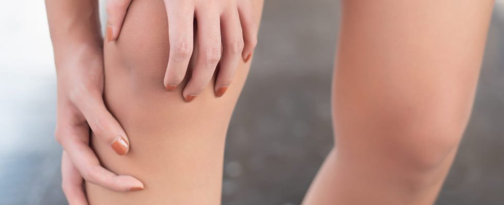
Erysipelas is a skin disease. It exists in two forms, a classic shape at the level of the face and a shape at the level of a inferior member. Erysipelas of the face results in a circle-shaped redness, associated with a sudden onset fever. Erysipelas inferior member is what we call the big leg syndrome, which is red, hot and which presents a edema. It’s a medical emergency and requires going to the hospital emergency room.
A streptococcal infection
Erysipelas is a bacterial infection of the skin, most commonly caused by a streptococcus. This disease is not superficial, since it affects the second layer of skin which is the dermis. “It’s a deep infection, don’tt the incidence is decreasing in France. explains Doctor Roland Viraben, dermatologist and member of the National Union of Dermatologists and Venereologists (SNDV). Erysipelas is more linked to hypersensitivity to the bacteria than the bacteria itself. Furthermore, the streptococcal infectionsin general, are much less frequent than before, in particular thanks to antibiotic therapies and, among other things, the best dental and ENT treatments.”
Venous insufficiency
Venous insufficiency is a risk factor in the occurrence of erysipelas. “The main cause of erysipelas is poor venous return, which is often hereditary. This poor venous circulation sensitizes the patient to infections, especially when its venous network is very altered. This state favors the appearance of venous stasis; the blood stagnates in the vein, thus creating local edema of the leg.
Foot fungus
“The second favoring element is the presence of a cutaneous portal of entry.” Indeed, the bacteria enters the skin via a wound, break or micro-fissure in the skin. “Often the bacteria enters the skin through small cracks between the toes, which are caused by fungus. Foot fungus is most often caused by poor foot hygiene or excess humidity and heat..”
A wound on the skin
For erysipelas to appear, a skin entry point is required. This is one of the modes of action of a bacteria: a minimal wound, a perleche, an ulcer… The bacteria takes advantage of this to pass through the altered skin barrier, which loses its protective role. Moreover, the search for this bacterial entry point must be systematically sought when treating erysipelas. If it is not found and treated with antibiotics, the risk of recurrence and complications is increased.
Cancer ?
No, erysipelas is not a consequence of cancer. “We never look for cancer in front of erysipelas. However, we are looking for the possible presence of a venous circulation disorder as well as cracks between the toes. Behind erysipelas on the lower limbwe absolutely must know if the patient is suffering from phlebitis“, reports our interlocutor. However, other general factors can influence the appearance of erysipelas, such as diabetes, obesity, alcoholism, smoking, immunodeficiency or even multiple antibiotic therapy.
Eczema or psoriasis?
Eczema and psoriasis are not the real causes of erysipelas. But in practice, these chronic dermatitis can cause skin breaks. It is through these gaps that the bacteria manages to penetrate into the dermis. “One of the only skin diseases that can possibly cause erysipelas is varicose eczema, but these are isolated and atypical cases“, indicates Dr Viraben. And as mentioned previously, foot fungus is likely to lead to micro-cracks, which are ultimately the entry point for the bacteria.
Treatment of erysipelas consists of administering antibiotics, under drip in case of fever. Then, an antibiotic treatment in the form of tablets, to be taken orally, is prescribed for a period of 3 weeks. Venous compression of the leg is proposed when possible to avoid peripheral venous insufficiency. Anti-inflammatories should be avoided because they promote infection. Anticoagulants may also be part of the treatment, only in specific cases. “These are only prescribed if there is associated phlebitis. Doppler ultrasound plethysmography allows the diagnosis of this possible complication.“, concludes the dermatologist.
Thanks to Doctor Roland Viraben, dermatologist and member of the National Union of Dermatologists and Venereologists (SNDV).
