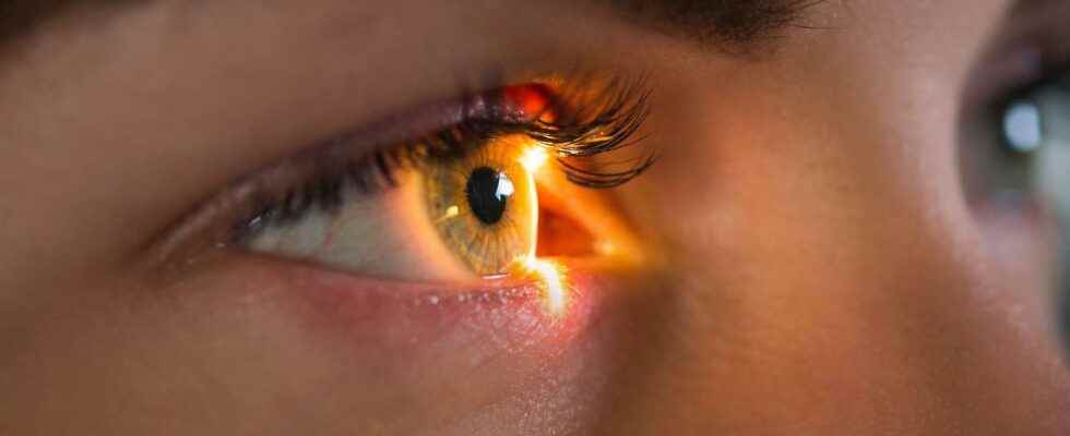
Serious diseases or common vision defects, the eyes are often affected by failures. Anti-color blindness glasses or falling asleep at the wheel, artificial cornea or gene therapy, here are the latest advances in eye health.
You will also be interested
[EN VIDÉO] Kezako: can we really trust our eyes? The human eye can differentiate nearly eight million shades of color. However, this organ so advanced gives little information to our cortex to create an image. So what exactly happens when we see? Unisciel and the University of Lille 1 explain to us, with the Kézako program, how this surprising body works.
About 80% of the information processed by the brain come from the eyes. This is to say if the health of the eyes is essential. However, many diseases affect vision, such as AMD (macular degeneration age-related), neuropathy hereditary by Leber, glaucoma, but also more common conditions such as myopia or color blindness. New treatments and innovative therapeutic devices promise to solve all our sight problems.
Stem cells to treat AMD
AMD is the leading cause of blindness after 50 years. Its “wet” version, the most common, is linked to an abnormal development of blood vessels which allows blood to leak. serum or blood in the retina and results in a loss of vision. Researchers from London Project to Cure Blindness (London project to cure blindness) have succeeded in partially restore vision in patients using embryonic stem cells developed on cells of theepithelium retinal. This tissue was then transplanted into the patients who were able to decipher and read words.
Glasses that curb myopia
Myopia affects a quarter of the world’s population and patients with high myopia are at increased risk of cataract and of retinal detachment. Reducing the impact of myopia is therefore a real public health issue. Japanese Hoya developed a spectacle lens incorporating tiny triangles at the periphery. These generate two distinct streams of images, which arrive in front and behind the retina. the brain treating the two streams as one, the elongation signaleye is blocked. According to the company, the progression of myopia in children is thus slowed down by 60% on average.
Glasses against falling asleep at the wheel
Some 25% of accidents of the road mortals are related to drowsiness. The startup Ellcie Healthy has designed frames with 15 sensors who record the signs of falling asleep (yawning, head micro-falls, frequency of eyelid closure ..). At the first signs of fatigue, the glasses send an alert to the wearer, in the form of a flashing or audible signal. Other occupants of the vehicle may also receive a signal on their smartphone. In the same vein, the startup has launched fall protection frames for the elderly, and trials are underway to detect seizures. Parkinson’s or to assess the shocks suffered during sport.
An artificial cornea to regain sight
When the cornea is damaged, the only possible solution is to have a new cornea transplanted from a donor. But in January 2021, 78-year-old man regained his sight thanks to an entirely artificial cornea. Consisting of a material porous non-degradable, it mimics the structure of extracellular matrix in order to provide support to the surrounding cells and to promote their proliferation. According to the Israeli company CorNeat Vision, which is behind it, theimplant is thus integrated in a few weeks. Another team of researchers succeeded in custom 4D printing of corneas that “mold” around the eye.
Optogenetics to activate the retina
THE’optogenetic consists of introducing into a cell a uncomfortable coding for a protein photosensitive, which will activate when it is illuminated with a light specific. A man with retinopathy pigmentary at an advanced stage (disease related to degeneration of light receptors in the retina) was thus able to partially regain sight, thanks to a gene sensitive to amber light introduced into retinal cells. When equipped with glasses that activate a certain type of light, its modified cells send a signal to the cortex visual allowing him to distinguish the shape and contours of objects.
Glasses against color blindness
An inherited male disease, color blindness affects about 8% of men. Caused by a deficiency of cones of the eye, photoreceptors located in the retina, it prevents us from distinguishing certain colors, such as red, orange, yellow, brown and green. Californian startup EnChroma invented glasses which selectively filter wavelengths at the border between red and green, where the confusion of colors occurs. In August 2020, researchers from theUC Davis Eye Center (United States) and Inserm have shown positive results for these glasses, the brain learning to detect small color variations such as different colors.
Improve sleep or relieve migraine
The eye does not only act on vision but also on health in general. We know, for example, that exposure to light promotes the production of melatonin, which makes it easier to fall asleep. By developing specific filters, it would thus be possible to act on the production of this hormone. Likewise, certain wavelengths activate photosensitive cells which worsen the pain linked to headache. The manufacturer Avalux has thus developed migraine glasses that filter this type of light (in the spectrum of blue light, amber and red) while letting in the soothing green light.
The iris, the colored part of the eye The iris is the colored area of the eye, visible at the front. It is in fact the anterior part of the vascular tunic (choroid). The iris has an opening in its center: the pupil, round, which lets light through. The diameter of the pupil can vary, allowing more or less light to enter, depending on the ambient light. The iris contains a more or less abundant brown pigment. © Mattis2412, CC by-sa 3.0
The lateral rectus muscle turns the eye The lateral rectus muscle of the eye rotates this organ around an axis, thus allowing the gaze to shift outwards. © Patrick J. Lynch, CC by 2.5
The consultation with the ophthalmologist During an examination at the ophthalmologist, the professional uses different lenses to test the patient’s eyesight. The measurement of the refractive error of the eye makes it possible to determine the prescription of glasses necessary for a good correction. © Nordelch, DP
Oblique muscle and trochlea of the eye This view of the anatomy of the eye makes it possible to visualize the trochlea: the oblique muscle, which has the same origin as the right muscles, forms a right angle while passing into the trochlea (top left), a kind of ” pulley »fibro-cartilaginous. © Patrick J. Lynch, CC by 2.5
Diagram of the eye: internal organization and envelopes of the eyeball The eyeball comprises three nested envelopes: a fibrous tunic: the sclera, or sclera (at the front, it is transparent and forms the cornea); a vascular tunic: the choroid; an internal tunic: the retina. Light enters the eye through the pupil, in the center of the iris. The light focuses on the retina, at the back of the eye, at the level of the macula (or yellow spot), where the concentration of cones is maximum. © Chabacano, CC by-sa 3.0
Diagram of a cross section of the eye In this diagram of a cross section of the eye, we see the lens, which is the lens of this sensory organ. It separates the eye into an anterior segment (at the front) and a posterior segment (at the back). The eye has transparent media: lens; aqueous humor before the lens; vitreous humor after the lens. The cornea is the part of the transparent sclera (or sclera) located at the front of the eye. © Rhcastilhos, DP
The tear system The lacrimal system consists of: a lacrimal gland; ducts. The lacrimal gland secretes saline (tears), which contains mucus, antibodies and an antibacterial enzyme called “lysozyme”. The ducts carry tear fluid. The blinking of the eyes pushes the tears down. © FML, CC by-sa 2.5
Muscles of the eye seen from the front Different muscles allow the eye to move: the upper rectus muscle; the lower rectus muscle; the lateral rectus muscle; the medial rectus muscle; the inferior oblique muscle; the superior oblique muscle. The lower oblique muscle helps, for example, to move the eye upwards. These muscles are innervated. © Patrick J. Lynch, CC by 2.5
The white of the eye From the front, the eye presents its colored iris which has an opening in the center, the pupil, which allows light to pass into the eye. Around the iris, the white of the eye is poorly vascularized. © Patrick J. Lynch, CC by 2.5
The human eye and additive synthesis The principle of additive synthesis is to reconstitute the appearance of colors by adding, in certain proportions, lights from three monochromatic sources. These three primary colors in the human eye are, depending on the wavelengths used by the cones of the retina: red; blue; green. © gt
Normal left fundus The fundus is the posterior wall of the retina. It is observed in ophthalmology consultation, thanks to the ophthalmoscope. This allows you to observe the blood vessels that lead from the optic nerve disc. © Mikael Häggström, CC0 1.0
Eye movements and optic nerve Eye movements are made possible by the muscles of the bulb of the eye, allowing the eye to follow the movement of objects. The optic nerve transmits visual information to the cortex. © Patrick J. Lynch, CC by 2.5
Interested in what you just read?
Subscribe to the newsletter The health question of the week : our answer to a question you ask yourself (more or less secretly). All our newsletters
.
fs7
