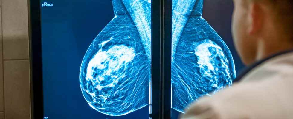
Breast calcifications are calcium accumulations in the tissues that make up the breast. Their presence is classically demonstrated after a mammogram (X-ray of the breasts). Most are not associated with cancer, but may reveal cancer.
Definition: what is breast calcification?
Breast calcifications are calcium deposits that form in the breast tissue. They have no connection with the amount of calcium absorbed during the diet or obtained through food supplements. “Breast calcifications are quite common and most are benign after age 40explains Dr. Elisabeth Paganelli, medical gynecologist. Their presence is highlighted after a breast x-ray“. But they may be associated with cancer. “In order to make a diagnosis, the radiologist studies the mammogram and on the images he looks at the small white dots, he assesses their size, shape and arrangement.“. If necessary, he takes additional shots to study these very small images in greater detail.
We will thus distinguish two types of breast calcifications:
► “Macrocalcificationsgenerally considered non-cancerous, are associated with aging of the mammary arteries or the existence of a benign injury or tumor“. They are found in about half of women over 50 and one in 10 women under 50.
► “And the microcalcificationswhich may be a sign of precancerous cells or early-stage breast cancer“.
What are the symptoms of breast calcification?
“Asymptomatic breast calcifications are not responsible for any symptoms and are too small to be felt during a breast palpationcontinues the gynecologist. Usually they are diagnosed on a mammogram in the form of small white marks“.”However, they may be visible in association with lesions that are palpable, whether benign or malignant nodules“, adds Dr. Caroline Malhaire, radiologist.
What are the causes of breast calcifications?
Breast calcifications are more common with aging and almost always occur after age 50. “The mechanisms of formation of calcifications are multiplecontinues Dr. Malhaire : aging of the vessels whose walls become calcified, calcium deposits in nodules or cysts, or scars from surgery or trauma to the breast, among others, for mild cases. The proliferation of atypical or cancerous cells can lead to the formation of calcification deposits and thus reveal a beginning lesion“. The search for abnormalities in mammography to diagnose and treat cancer early is the principle of organized screening for breast cancer in France.
50% of breast cancer cases are associated with breast calcifications
Is this a sign of cancer?
We think that 50% of breast cancer cases are associated with breast calcifications. Suspicion will be high if the doctor finds irregular calcifications, or in a dense spiculated area. “Analysis of the area of the breast that contains microcalcifications can be done using a biopsy system guided by mammography, which will allow microscopic analysis of breast tissue fragments containing these calcifications and to define their benign, atypical or malignant origin“, adds the radiologist.
When and who to consult in case of breast calcifications?
“In general it is the radiologist reading the mammogramwhich performs the diagnosis and offers you a course of action according to recommendations: simple monitoring, delay, rapid sampling for analysis of the suspect area…”, recalls the gynecologist. The patient can consult her general practitioner or gynecologist with his shots.
“Depending on the type of microcalcifications, we propose simple monitoring, or close examinationsanswers Dr. Paganelli. If the area of microcalcifications is suspicious, the radiologist will offer to quickly perform a biopsy in order to check the sampled tissue which will be entrusted to and studied by a pathologist“. According to the Bi-rads classification (Breast Imaging Reporting And Data System) of the American College of Radiology, we differentiate for example:
► An ACR3-classified mammogram (or Bi-rads 3), corresponding to a mammogram with a most likely benign abnormality (>97% of benign lesions in the literature), for which monitoring is recommended at 6, 12 and 24 months. “If there are no changes during this close monitoring, the patient can resume her usual follow-up“, says Dr. Malhaire.
► An ACR4-classified mammogram (or Bi-rads 4), corresponds to a mammogram with an indeterminate or suspicious abnormality, for which further investigations are necessary (mainly mammography-guided macrobiopsies). Often a subdivision into ACR4a, ACR4b, and ACR4c is performed to better assess the risk of malignancy.
► An ACR5-classified mammogram (or Bi-rads 5), corresponds to a mammogram with an anomaly strongly suggestive of breast cancer, and for which the continuation of the investigations by biopsies is essential.
What is the treatment for removing breast calcifications?
Breast calcifications are not a disease or disorder in themselves. They therefore do not require treatment as such. “It is the underlying abnormality they reveal that may require treatment. in a specialized senology service in the event of cancer, close monitoring, or no change in care if the biopsies ultimately show benign lesions“, insists the radiologist. The measures to be taken depend on the imaging and the pathological result of the sample if it took place. “If the analysis of the area of breast tissue that contains breast microcalcifications finds cancerous cells, the patient will be taken care of for her cancer after a collegial decision in a Multidisciplinary Consultation Meeting according to national recommendations (localized surgery, chemotherapy, radiotherapy, hormone therapy, mastectomy, etc.)“, concludes Dr. Paganelli.
Thanks to Dr Caroline Malhaire, Radiologist at the Institut Curie and Dr Elisabeth Paganelli, Medical Gynecologist and General Secretary of Syngof (union of medical gynecologists and obstetrician gynecologists)
